SKU: 13-2024
Size/Quantity: 100 ul
SP1 CUTANA™ CUT&RUN Antibody
$545.00
In stock
 Powered by Bioz


In stock
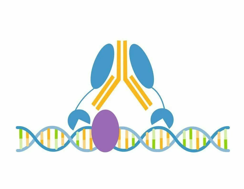
This antibody meets EpiCypher’s “CUTANA Compatible” criteria for performance in Cleavage Under Targets and Release Using Nuclease (CUT&RUN) and/or Cleavage Under Targets and Tagmentation (CUT&Tag) approaches to genomic mapping. Every lot of a CUTANA Compatible antibody is tested in the indicated approach using EpiCypher optimized protocols and determined to yield peaks that show a genomic distribution pattern consistent with reported function(s) of the target protein. SP1 is the original member of the Sp1-like family of zinc-finger transcription factors. It binds GC-rich motifs with high affinity [1] and is involved in expressing genes related to cell growth, cell-cycle progression, survival, and tumorigenesis [2]. SP1 antibody produces CUT&RUN peaks above background primarily in intronic and promoter regions (Figure 1) that overlap with known SP1 DNA-binding motifs (Figure 2).
CUT&RUN was performed on 500k HeLa cells with 0.5 µg of either SP1 or IgG negative control (EpiCypher 13-0042) antibodies using the CUTANA™ ChIC/CUT&RUN Kit v2.0 (EpiCypher 14-1048). Library preparation was performed with 5 ng of DNA (or the total amount recovered if less than 5 ng) using the CUTANA™ CUT&RUN Library Prep Kit (EpiCypher 14-1001/14-1002). Both kit protocols were adapted for high throughput Tecan liquid handling. Libraries were run on an Illumina NextSeq2000 with paired-end sequencing (2×50 bp). Sample sequencing depth was 5.0 million reads (IgG) and 5.3 million reads (SP1). Data were aligned to the hg19 genome using Bowtie2. Data were filtered to remove duplicates, multi-aligned reads, and ENCODE DAC Exclusion List regions.