SKU: 13-2013
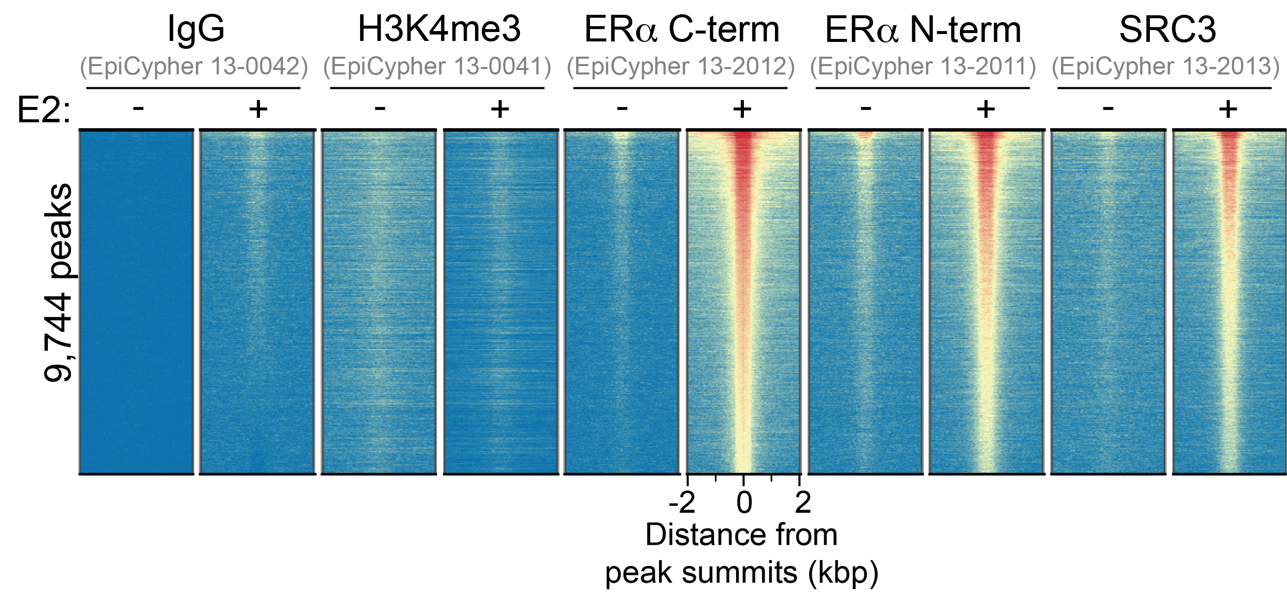
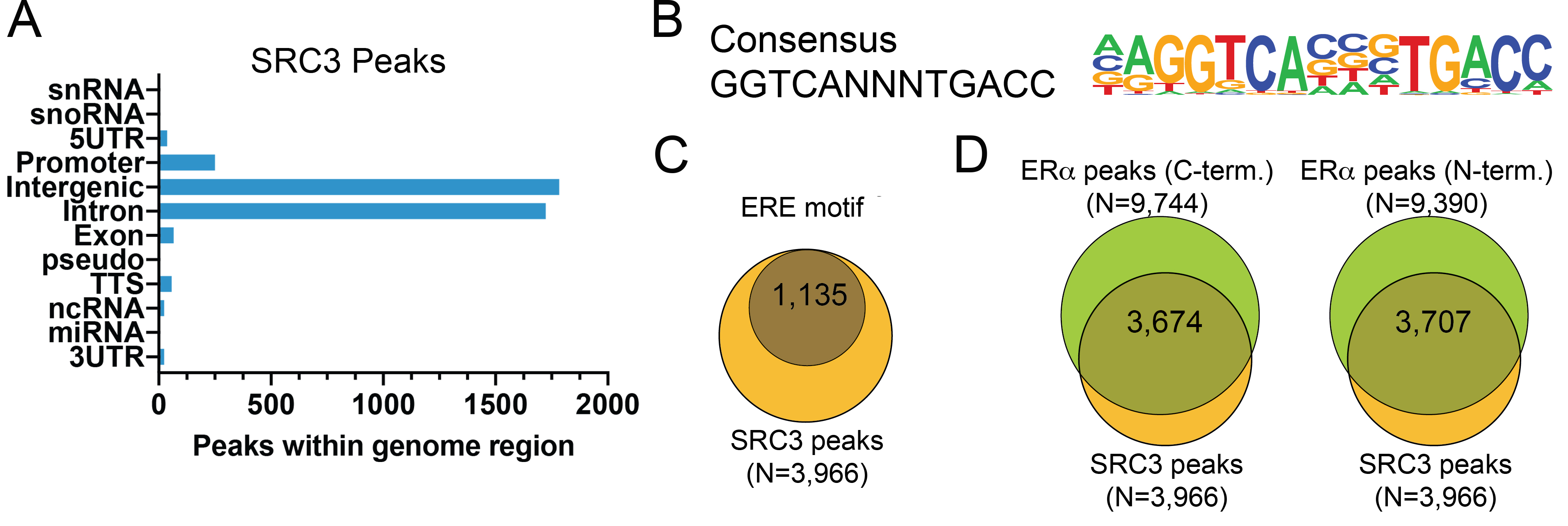
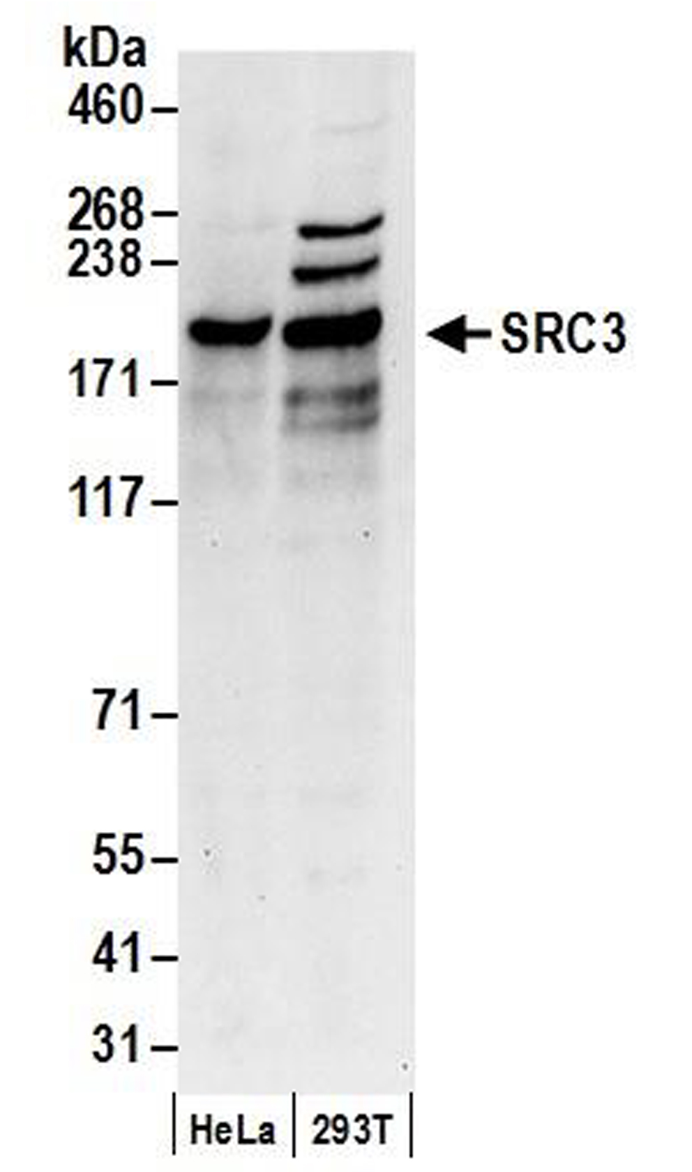
{"url":"https://www.epicypher.com/products/antibodies/cutana-cut-run-antibodies/ncoa3-src3-cutana-cut-run-antibody","add_this":[{"service":"facebook","annotation":""},{"service":"email","annotation":""},{"service":"print","annotation":""},{"service":"twitter","annotation":""},{"service":"linkedin","annotation":""}],"gtin":null,"id":842,"bulk_discount_rates":[],"can_purchase":true,"meta_description":"Polyclonal anti-NCOA3/SRC3 antibody validated for CUT&RUN, WB. Rigorously tested for reliable and robust performance in CUT&RUN assays.","category":["Epigenetics Kits and Reagents/CUTANA™ ChIC / CUT&RUN Assays","Antibodies/CUTANA™ CUT&RUN Antibodies","Antibodies/CUTANA™ CUT&RUN Antibodies/CUT&RUN Antibodies - Chromatin-Associated Proteins"],"AddThisServiceButtonMeta":"","main_image":{"data":"https://cdn11.bigcommerce.com/s-y9o92/images/stencil/{:size}/products/842/1005/ncoa3src3-cutana-cutandrun-antibody__47372.1645734559__68175.1648666372.jpg?c=2","alt":"NCOA3/SRC3 CUTANA™ CUT&RUN Antibody"},"add_to_wishlist_url":"/wishlist.php?action=add&product_id=842","shipping":{"calculated":true},"num_reviews":0,"weight":"0.00 LBS","custom_fields":[{"id":"713","name":"Pack size","value":"100 µL"}],"sku":"13-2013","description":"<div class=\"product-general-info\">\n <ul class=\"product-general-info__list-left\">\n <li class=\"product-general-info__list-item\">\n <strong>Type: </strong>Polyclonal\n </li>\n <li class=\"product-general-info__list-item\">\n <strong>Host: </strong>Rabbit\n </li>\n <li class=\"product-general-info__list-item\">\n <strong>Target Size: </strong>155 kDa\n </li>\n </ul>\n <ul class=\"product-general-info__list-right\">\n <li class=\"product-general-info__list-item\">\n <strong>Format: </strong>Antigen affinity-purified\n </li>\n <li class=\"product-general-info__list-item\">\n <strong>Reactivity: </strong>Human\n </li>\n <li class=\"product-general-info__list-item\">\n <strong>Applications: </strong>CUT&RUN, WB\n </li>\n </ul>\n</div>\n<div class=\"service_accordion product-droppdown\">\n <div class=\"container\">\n <div id=\"prodAccordion\">\n <div id=\"ProductDescription\" class=\"Block Panel current\">\n <h3 class=\"sub-title1\">Description</h3>\n <div\n class=\"ProductDescriptionContainer product-droppdown__section-description-specific\">\n <p>\n This antibody meets EpiCypher’s “CUTANA Compatible” criteria for\n performance in Cleavage Under Targets and Release Using Nuclease\n (CUT&RUN) and/or Cleavage Under Targets and Tagmentation\n (CUT&Tag) approaches to genomic mapping. Every lot of a CUTANA\n Compatible antibody is tested in the indicated CUTANA approach using\n <a href=\"https://www.epicypher.com/resources/protocols\"\n >EpiCypher optimized protocols\n </a>\n and determined to yield peaks that show a genomic distribution\n pattern consistent with reported function(s) of the target protein.\n Consistent with its role as a nuclear receptor coactivator that\n facilitates ER-mediated transcription (1), NCOA3 (SRC3) antibody\n shows CUT&RUN peaks in response to estradiol stimulation\n <strong>(Figure 1)</strong> that overlap with known estrogen\n response element (ERE) binding motifs <strong>(Figure 2)</strong>.\n SRC3 CUT&RUN peaks also overlap with ER alpha antibodies (N- and\n C-term;\n <a\n href=\"/products/antibodies/cutana-cut-run-compatible-antibodies/estrogen-receptor-alpha-n-terminal-cutana-cut-run-antibody\"\n >13-2011</a\n >\n and\n <a\n href=\"/products/antibodies/cutana-cut-run-compatible-antibodies/estrogen-receptor-alpha-c-terminal-cutana-cut-run-antibody\"\n >13-2012</a\n >\n respectively) (Figure 2).\n </p>\n </div>\n </div>\n </div>\n <div id=\"prodAccordion\">\n <div id=\"ProductDescription\" class=\"Block Panel current\">\n <h3 class=\"sub-title1\">Validation Data</h3>\n <div\n class=\"ProductDescriptionContainer product-droppdown__section-description-specific\">\n <section class=\"image-picker\">\n <div class=\"image-picker__left\">\n <div\n class=\"image-picker__main-content_active image-picker__main-content\">\n <div class=\"image-picker__header-content\">\n <button class=\"image-picker__left-arrow\">\n <svg\n class=\"image-picker__svg-left\"\n width=\"24\"\n height=\"24\"\n viewBox=\"0 0 24 24\">\n <path\n d=\"M16.67 0l2.83 2.829-9.339 9.175 9.339 9.167-2.83 2.829-12.17-11.996z\" />\n </svg>\n </button>\n <a\n href=\"/content/images/products/antibodies/13_2013_Heatmap.jpeg\"\n target=\"_new\"\n class=\"image-picker__main-image-link\"\n ><img loading=\"lazy\"\n alt=\"13_2013_Heatmap\"\n src=\"/content/images/products/antibodies/13_2013_Heatmap.jpeg\"\n class=\"image-picker__main-image\" />\n <span class=\"image-picker__main-image-caption\"\n >(Click to enlarge)</span\n ></a\n >\n <button class=\"image-picker__right-arrow\">\n <svg\n class=\"image-picker__svg-right\"\n width=\"24\"\n height=\"24\"\n viewBox=\"0 0 24 24\">\n <path\n d=\"M7.33 24l-2.83-2.829 9.339-9.175-9.339-9.167 2.83-2.829 12.17 11.996z\" />\n </svg>\n </button>\n </div>\n <p>\n <span class=\"image-picker__span-content\"\n ><strong>Figure 1: SRC3 enrichment in CUT&RUN </strong\n ><br />\n Serum-starved MCF7 cells were treated with 100 nM estradiol\n (E2) or vehicle control for 45 minutes. CUT&RUN was\n performed using 500,000 cells with 0.5 µg of each indicated\n antibody (gray text). Heatmap shows CUT&RUN enrichment in\n aligned rows ranked by intensity (top to bottom; relative to\n ER alpha C-term (<a\n href=\"/products/antibodies/cutana-cut-run-compatible-antibodies/estrogen-receptor-alpha-c-terminal-cutana-cut-run-antibody\"\n >13-2012</a\n >). Red indicates high localized enrichment and blue denotes\n background signal.\n </span>\n </p>\n </div>\n <div class=\"image-picker__main-content\">\n <div class=\"image-picker__header-content\">\n <button class=\"image-picker__left-arrow\">\n <svg\n class=\"image-picker__svg-left\"\n width=\"24\"\n height=\"24\"\n viewBox=\"0 0 24 24\">\n <path\n d=\"M16.67 0l2.83 2.829-9.339 9.175 9.339 9.167-2.83 2.829-12.17-11.996z\" />\n </svg>\n </button>\n <a\n href=\"/content/images/products/antibodies/13_2013_Peaks.jpeg\"\n target=\"_new\"\n class=\"image-picker__main-image-link\"\n ><img loading=\"lazy\"\n alt=\"13_2013_Peaks\"\n src=\"/content/images/products/antibodies/13_2013_Peaks.jpeg\"\n class=\"image-picker__main-image\" />\n <span class=\"image-picker__main-image-caption\"\n >(Click to enlarge)</span\n ></a\n >\n <button class=\"image-picker__right-arrow\">\n <svg\n class=\"image-picker__svg-right\"\n width=\"24\"\n height=\"24\"\n viewBox=\"0 0 24 24\">\n <path\n d=\"M7.33 24l-2.83-2.829 9.339-9.175-9.339-9.167 2.83-2.829 12.17 11.996z\" />\n </svg>\n </button>\n </div>\n <p>\n <span class=\"image-picker__span-content\"\n ><strong>Figure 2: SRC3 peak analysis in CUT&RUN</strong\n ><br />\n Peaks from the E2- treated samples in Figure 1 were called\n using MACS2. <strong>(A)</strong> The number of SRC3 peaks\n which fall into distinct classes of functionally annotated\n genomic regions is plotted. <strong>(B)</strong> Homer\n analysis determined that the ERE consensus motif,\n represented as a sequence logo position weight matrix, was\n enriched under SRC3 peaks. <strong>(C)</strong> The number\n of SRC3 peaks containing consensus motifs from panel B is\n shown by Venn Diagram. <strong>(D)</strong> The number of\n SRC3 peaks that overlap with ER alpha C-term (<a\n href=\"/products/antibodies/cutana-cut-run-compatible-antibodies/estrogen-receptor-alpha-c-terminal-cutana-cut-run-antibody\"\n >13-2012</a\n >) and ER alpha N-term (<a\n href=\"/products/antibodies/cutana-cut-run-compatible-antibodies/estrogen-receptor-alpha-n-terminal-cutana-cut-run-antibody\"\n >13-2011</a\n >) antibodies are represented by Venn Diagram.\n </span>\n </p>\n </div>\n <div class=\"image-picker__main-content\">\n <div class=\"image-picker__header-content\">\n <button class=\"image-picker__left-arrow\">\n <svg\n class=\"image-picker__svg-left\"\n width=\"24\"\n height=\"24\"\n viewBox=\"0 0 24 24\">\n <path\n d=\"M16.67 0l2.83 2.829-9.339 9.175 9.339 9.167-2.83 2.829-12.17-11.996z\" />\n </svg>\n </button>\n <a\n href=\"/content/images/products/antibodies/13_2013_WB.jpeg\"\n target=\"_new\"\n class=\"image-picker__main-image-link\"\n ><img loading=\"lazy\"\n alt=\"13_2013_WB\"\n src=\"/content/images/products/antibodies/13_2013_WB.jpeg\"\n class=\"image-picker__main-image\" />\n <span class=\"image-picker__main-image-caption\"\n >(Click to enlarge)</span\n ></a\n >\n <button class=\"image-picker__right-arrow\">\n <svg\n class=\"image-picker__svg-right\"\n width=\"24\"\n height=\"24\"\n viewBox=\"0 0 24 24\">\n <path\n d=\"M7.33 24l-2.83-2.829 9.339-9.175-9.339-9.167 2.83-2.829 12.17 11.996z\" />\n </svg>\n </button>\n </div>\n <p>\n <span class=\"image-picker__span-content\"\n ><strong\n >Figure 3: Western blot detection of human SRC3</strong\n ><br />\n Whole cell lysates were isolated from HeLa and HEK293T cells\n using NETN lysis buffer. Lysate (50 µg) was loaded onto 4-8%\n SDS-PAGE gel and analyzed under standard western blot\n conditions using SRC3 antibody (0.1 µg/mL).\n </span>\n </p>\n </div>\n </div>\n <aside class=\"image-picker__right\">\n <div class=\"image-picker__gallery\">\n <img loading=\"lazy\"\n alt=\"13_2009_Heatmap\"\n src=\"/content/images/products/antibodies/13_2013_Heatmap.jpeg\"\n width=\"200\"\n class=\"image-picker__side-image image-picker__side-image_active\"\n role=\"button\" />\n <img loading=\"lazy\"\n alt=\"13_2013_Peaks\"\n src=\"/content/images/products/antibodies/13_2013_Peaks.jpeg\"\n class=\"image-picker__side-image\"\n role=\"button\" />\n <img loading=\"lazy\"\n alt=\"13_2013_WB\"\n src=\"/content/images/products/antibodies/13_2013_WB.jpeg\"\n class=\"image-picker__side-image\"\n role=\"button\" />\n </div>\n </aside>\n </section>\n </div>\n </div>\n </div>\n <div id=\"prodAccordion\">\n <div id=\"ProductDescription\" class=\"Block Panel\">\n <h3 class=\"sub-title1\">Technical Information</h3>\n <div\n class=\"ProductDescriptionContainer product-droppdown__section-description\">\n <div class=\"product-tech-info\">\n <div class=\"product-tech-info__line-item\">\n <div class=\"product-tech-info__line-item-left\">\n <b>Immunogen</b>\n </div>\n\n <div class=\"product-tech-info__line-item-right\">\n A synthetic peptide corresponding to human NCOA3/SRC3 amino\n acids 900 - 950.\n </div>\n </div>\n <div class=\"product-tech-info__line-item\">\n <div class=\"product-tech-info__line-item-left\">\n <b>Formulation</b>\n </div>\n <div class=\"product-tech-info__line-item-right\">\n Antigen affinity-purified antibody (1.0 mg/mL) in\n Tris-citrate/phosphate buffer pH 7 to 8, 0.09% sodium azide.\n </div>\n </div>\n <div class=\"product-tech-info__line-item\">\n <div class=\"product-tech-info__line-item-left\">\n <b>Storage and Stability</b>\n </div>\n <div class=\"product-tech-info__line-item-right\">\n Stable for 1 year at 4°C from date of receipt.\n </div>\n </div>\n </div>\n </div>\n </div>\n </div>\n <div id=\"prodAccordion\">\n <div id=\"ProductDescription\" class=\"Block Panel\">\n <h3 class=\"sub-title1\">Application Notes</h3>\n <div\n class=\"ProductDescriptionContainer product-droppdown__section-description\">\n <p><strong>Recommended Dilutions:</strong></p>\n <p><strong>CUT&RUN:</strong> 0.5 µg</p>\n <p><strong>WB:</strong> 1:2000 - 1:10,000</p>\n </div>\n </div>\n </div>\n <div id=\"prodAccordion\">\n <div id=\"ProductDescription\" class=\"Block Panel\">\n <h3 class=\"sub-title1\">References</h3>\n <div\n class=\"ProductDescriptionContainer product-droppdown__section-description\">\n <strong>Background References:</strong>\n <br />\n 1. Wagner <em>et al.</em> <em>BMC Cancer </em> (2013). PMID:\n <a\n href=\"https://pubmed.ncbi.nlm.nih.gov/24304549/\"\n title=\"NCOA3 is a selective co-activator of estrogen receptor α-mediated transactivation of PLAC1 in MCF-7 breast cancer cells\"\n target=\"new\">\n 24304549</a\n ><br />\n </div>\n </div>\n </div>\n <div id=\"prodAccordion\">\n <div id=\"ProductDescription\" class=\"Block Panel\">\n <h3 class=\"sub-title1\">Documents & Resources</h3>\n <div\n class=\"ProductDescriptionContainer product-droppdown__section-description\">\n <div class=\"product-documents\">\n <a\n href=\"/content/documents/tds/13-2013.pdf\"\n target=\"_new\"\n class=\"product-documents__link\">\n <svg\n version=\"1.1\"\n id=\"Layer_1\"\n xmlns=\"http://www.w3.org/2000/svg\"\n xmlns:xlink=\"http://www.w3.org/1999/xlink\"\n x=\"0px\"\n y=\"0px\"\n viewBox=\"0 0 228 240\"\n style=\"enable-background: new 0 0 228 240\"\n xml:space=\"preserve\"\n class=\"product-documents__icon\">\n <g>\n <path\n class=\"product-documents__svg-pdf\"\n d=\"M191.92,68.77l-47.69-47.69c-1.33-1.33-3.12-2.08-5.01-2.08H45.09C41.17,19,38,22.17,38,26.09v184.36\n c0,3.92,3.17,7.09,7.09,7.09h141.82c3.92,0,7.09-3.17,7.09-7.09V73.8C194,71.92,193.25,70.1,191.92,68.77z M177.65,77.06h-41.7\n v-41.7L177.65,77.06z M178.05,201.59H53.95V34.95h66.92v47.86c0,5.14,4.17,9.31,9.31,9.31h47.86V201.59z\" />\n </g>\n <rect\n x=\"20\"\n y=\"112\"\n class=\"product-documents__svg-background\"\n width=\"146\"\n height=\"76\" />\n <g>\n <path\n class=\"product-documents__svg-pdf\"\n d=\"M23.83,125.68h22.36c5.29,0,9.41,1.33,12.35,4c2.94,2.67,4.42,6.39,4.42,11.18c0,4.78-1.47,8.51-4.42,11.18\n c-2.94,2.67-7.06,4-12.35,4H34.59v18.29H23.83V125.68z M44.81,147.9c5.38,0,8.07-2.32,8.07-6.97c0-2.39-0.67-4.16-2-5.31\n c-1.33-1.15-3.36-1.73-6.07-1.73H34.59v14.01H44.81z\" />\n <path\n class=\"product-documents__svg-pdf\"\n d=\"M69.92,125.68h18.91c5.29,0,9.84,0.97,13.66,2.9c3.82,1.93,6.74,4.72,8.76,8.35\n c2.02,3.63,3.04,7.98,3.04,13.04c0,5.06-1,9.42-3,13.08c-2,3.66-4.91,6.45-8.73,8.38c-3.82,1.93-8.4,2.9-13.73,2.9H69.92V125.68z\n M88.07,165.63c10.35,0,15.52-5.22,15.52-15.66c0-10.4-5.17-15.59-15.52-15.59h-7.38v31.26H88.07z\" />\n <path\n class=\"product-documents__svg-pdf\"\n d=\"M122.57,125.68h32.84v8.49h-22.22v11.18h20.84v8.49h-20.84v20.49h-10.63V125.68z\" />\n </g>\n </svg>\n <span class=\"product-documents__info\">Technical Datasheet</span>\n </a>\n </div>\n </div>\n </div>\n </div>\n <div id=\"prodAccordion\">\n <div id=\"ProductDescription\" class=\"Block Panel\">\n <h3 class=\"sub-title1\">Additional Info</h3>\n <div\n class=\"ProductDescriptionContainer product-droppdown__section-description\">\n <p>\n This product is provided for commercial sale under license from\n Bethyl Laboratories, Inc.\n </p>\n\n <strong>Applications Key:</strong> <br /><br />\n\n ChIP: Chromatin immunoprecipitation<br />\n CUT&RUN: Cleavage Under Targets and Release Using Nuclease<br />\n CUT&Tag: Cleavage Under Targets and Tagmentation<br />\n E: ELISA<br />\n FACS: Flow cytometry<br />\n ICC: Immunocytochemistry<br />\n IF: Immunofluorescence<br />\n IHC: Immunohistochemistry<br />\n IP: Immunoprecipitation<br />\n L: Luminex<br />\n WB: Western Blot<br /><br />\n\n <strong>Reactivity Key:</strong> <br /><br />\n\n B: Bovine<br />\n Ce: <em>C. elegans</em><br />\n Ch: Chicken<br />\n Dm: <em>Drosophila</em><br />\n Eu: Eukaryote<br />\n H: Human<br />\n M: Mouse<br />\n Ma: Mammal<br />\n R: Rat<br />\n Sc: <em>S. cerevisiae</em><br />\n Sp: <em>S. pombe</em><br />\n WR: Wide Range (predicted)<br />\n X: Xenopus<br />\n Z: Zebrafish<br />\n </div>\n </div>\n </div>\n </div>\n</div>\n","tags":[],"warranty":"","price":{"without_tax":{"formatted":"$525.00","value":525,"currency":"USD"},"tax_label":"Sales Tax"},"detail_messages":"","availability":"","page_title":"NCOA3/SRC3 Antibody | CUTANA™ CUT&RUN Compatible","cart_url":"https://www.epicypher.com/cart.php","max_purchase_quantity":0,"mpn":null,"upc":null,"options":[],"related_products":[{"id":889,"sku":null,"name":"CUTANA™ CUT&RUN Library Prep Kit","url":"https://www.epicypher.com/products/epigenetics-kits-and-reagents/cutana-cut-run-library-prep-kit","availability":"","rating":null,"brand":{"name":null},"category":["Epigenetics Kits and Reagents","Epigenetics Kits and Reagents/CUTANA™ ChIC / CUT&RUN Assays"],"summary":"\n \n \n \n \n \n \n \n ","image":{"data":"https://cdn11.bigcommerce.com/s-y9o92/images/stencil/{:size}/products/889/997/Epicypher_CUTANA_CUTRUN_LibraryPrep_Box__64431.1646406521.png?c=2","alt":"CUTANA™ CUT&RUN Library Prep Kit"},"images":[{"data":"https://cdn11.bigcommerce.com/s-y9o92/images/stencil/{:size}/products/889/997/Epicypher_CUTANA_CUTRUN_LibraryPrep_Box__64431.1646406521.png?c=2","alt":"CUTANA™ CUT&RUN Library Prep Kit"}],"date_added":"31st Jan 2022","pre_order":false,"show_cart_action":true,"has_options":true,"stock_level":null,"low_stock_level":null,"qty_in_cart":0,"custom_fields":[{"id":983,"name":"Pack Size","value":"48 Reactions"}],"num_reviews":null,"weight":{"formatted":"0.01 LBS","value":0.01},"demo":false,"price":{"without_tax":{"currency":"USD","formatted":"$1,525.00","value":1525},"tax_label":"Sales Tax"},"add_to_wishlist_url":"/wishlist.php?action=add&product_id=889"},{"id":694,"sku":null,"name":"CUTANA™ pAG-MNase for ChIC/CUT&RUN Workflows","url":"https://www.epicypher.com/products/epigenetics-reagents-and-assays/cutana-pag-mnase-for-chic-cut-and-run-workflows","availability":"","rating":null,"brand":{"name":null},"category":["Epigenetics Kits and Reagents","Epigenetics Kits and Reagents/CUTANA™ ChIC / CUT&RUN Assays"],"summary":"\n \n \n Type: Nuclease\n \n \n Mol Wgt: 43.7 kDa\n \n \n \n ","image":{"data":"https://cdn11.bigcommerce.com/s-y9o92/images/stencil/{:size}/products/694/689/Screen_Shot_2020-02-12_at_11.01.55_AM__17144.1581530752.png?c=2","alt":"CUTANA™ pAG-MNase for ChIC/CUT&RUN Workflows"},"images":[{"data":"https://cdn11.bigcommerce.com/s-y9o92/images/stencil/{:size}/products/694/689/Screen_Shot_2020-02-12_at_11.01.55_AM__17144.1581530752.png?c=2","alt":"CUTANA™ pAG-MNase for ChIC/CUT&RUN Workflows"}],"date_added":"12th Aug 2019","pre_order":false,"show_cart_action":true,"has_options":true,"stock_level":null,"low_stock_level":null,"qty_in_cart":0,"custom_fields":[{"id":1174,"name":"Internal Comment","value":"Excess in bottom of Venom"},{"id":1175,"name":"Internal Comment","value":"Bulk in Psylocke"}],"num_reviews":null,"weight":{"formatted":"0.01 LBS","value":0.01},"demo":false,"price":{"without_tax":{"currency":"USD","formatted":"$335.00","value":335},"price_range":{"min":{"without_tax":{"currency":"USD","formatted":"$335.00","value":335},"tax_label":"Sales Tax"},"max":{"without_tax":{"currency":"USD","formatted":"$1,295.00","value":1295},"tax_label":"Sales Tax"}},"tax_label":"Sales Tax"},"add_to_wishlist_url":"/wishlist.php?action=add&product_id=694"},{"id":758,"sku":"13-0042","name":"CUTANA™ IgG Negative Control Antibody for CUT&RUN and CUT&Tag","url":"https://www.epicypher.com/products/nucleosomes/snap-cutana-spike-in-controls/cutana-igg-negative-control-antibody-for-cut-run-and-cut-tag","availability":"","rating":null,"brand":{"name":null},"category":["Nucleosomes/SNAP-CUTANA™ Spike-in Controls","Antibodies/CUTANA™ CUT&RUN Antibodies","Epigenetics Kits and Reagents","Epigenetics Kits and Reagents/CUTANA™ ChIC / CUT&RUN Assays","Epigenetics Kits and Reagents/CUTANA™ CUT&Tag Assays"],"summary":"\n \n \n Type: Polyclonal\n \n \n Host: Rabbit\n \n \n Applications: CUT&R","image":{"data":"https://cdn11.bigcommerce.com/s-y9o92/images/stencil/{:size}/products/758/739/1579731877.1280.1280__61771.1592491196.png?c=2","alt":"CUTANA™ IgG Negative Control Antibody for CUT&RUN and CUT&Tag"},"images":[{"data":"https://cdn11.bigcommerce.com/s-y9o92/images/stencil/{:size}/products/758/739/1579731877.1280.1280__61771.1592491196.png?c=2","alt":"CUTANA™ IgG Negative Control Antibody for CUT&RUN and CUT&Tag"}],"date_added":"17th Jun 2020","pre_order":false,"show_cart_action":true,"has_options":false,"stock_level":null,"low_stock_level":null,"qty_in_cart":0,"custom_fields":[{"id":1148,"name":"Pack Size","value":"100 µg"},{"id":1149,"name":"Internal Comment","value":"bulk stocks from Thermo in cold room"}],"num_reviews":null,"weight":{"formatted":"0.01 LBS","value":0.01},"demo":false,"add_to_cart_url":"https://www.epicypher.com/cart.php?action=add&product_id=758","price":{"without_tax":{"currency":"USD","formatted":"$105.00","value":105},"tax_label":"Sales Tax"},"add_to_wishlist_url":"/wishlist.php?action=add&product_id=758"}],"shipping_messages":[],"rating":0,"meta_keywords":"ncoa3 antibody, src3 antibody, CUT&RUN antibody","show_quantity_input":1,"title":"NCOA3/SRC3 CUTANA™ CUT&RUN Antibody","gift_wrapping_available":false,"min_purchase_quantity":0,"customizations":[],"images":[{"data":"https://cdn11.bigcommerce.com/s-y9o92/images/stencil/{:size}/products/842/1005/ncoa3src3-cutana-cutandrun-antibody__47372.1645734559__68175.1648666372.jpg?c=2","alt":"NCOA3/SRC3 CUTANA™ CUT&RUN Antibody"},{"data":"https://cdn11.bigcommerce.com/s-y9o92/images/stencil/{:size}/products/842/967/ncoa3src3-cutana-cutandrun-antibody__47372.1645734559.jpg?c=2","alt":"NCOA3/SRC3 CUTANA CUTandRUN Antibody"}]}
Pack size: 100 µL