SKU: 13-0045
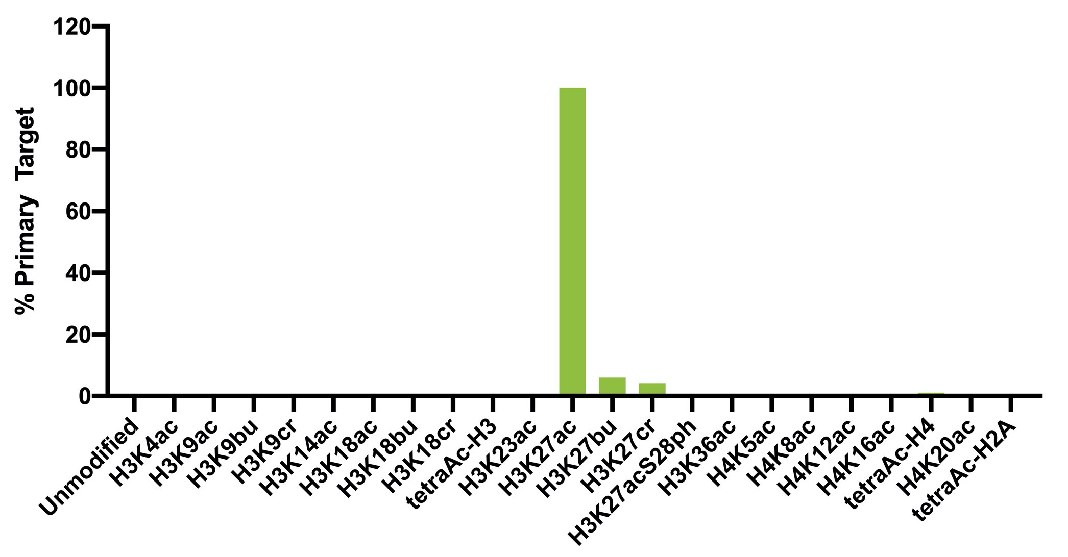
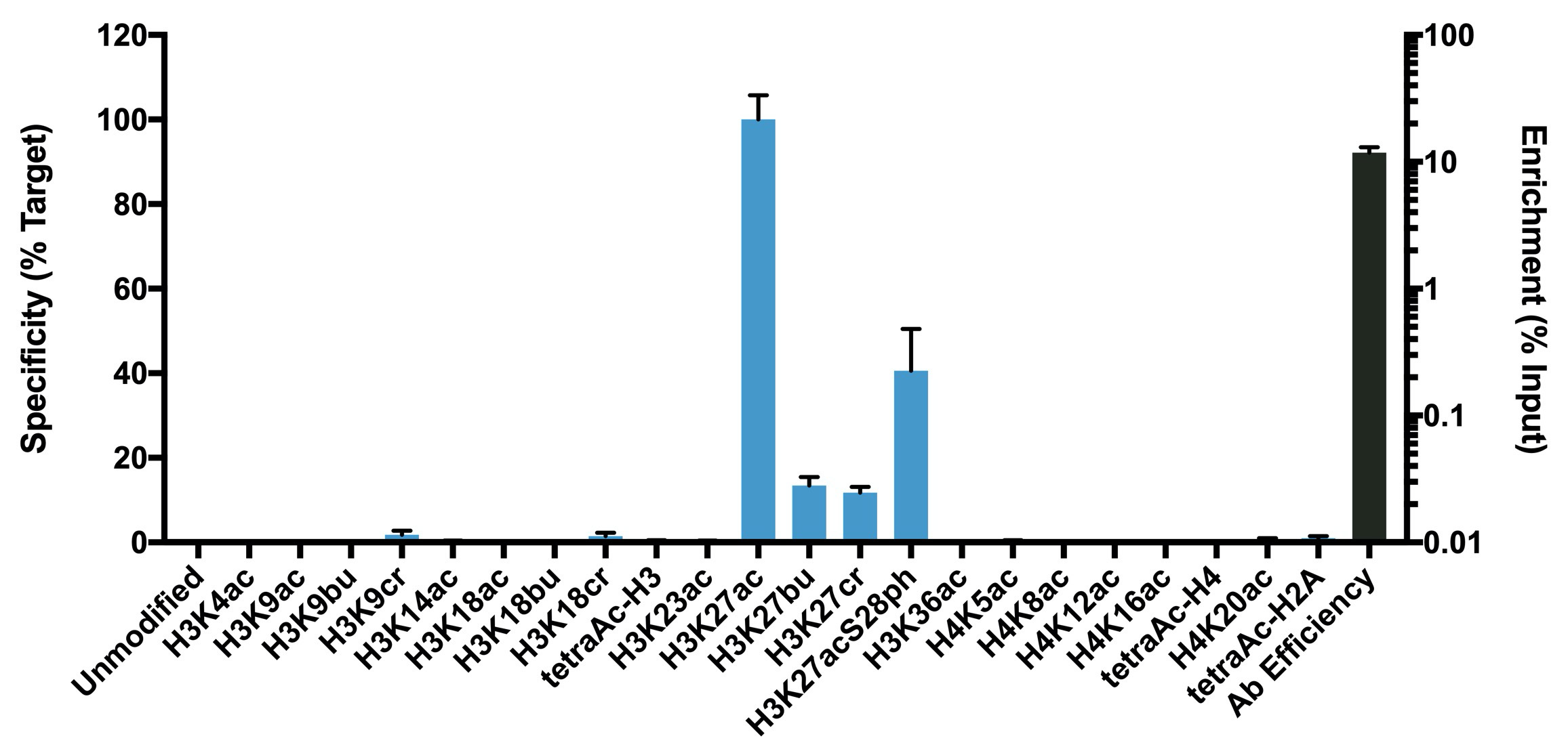
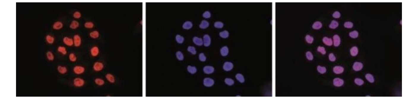
{"url":"https://www.epicypher.com/products/antibodies/snap-chip-certified-antibodies/histone-h3k27ac-antibody-snap-chip-certified","add_this":[{"service":"facebook","annotation":""},{"service":"email","annotation":""},{"service":"print","annotation":""},{"service":"twitter","annotation":""},{"service":"linkedin","annotation":""}],"warranty":"","gtin":null,"max_purchase_quantity":0,"id":759,"can_purchase":false,"meta_description":"Mouse monoclonal histone H3K27ac antibody rigorously tested for robust and reliable performance in ChIP assays. Also validated for ICC/IF.","category":["Discontinued Products"],"meta_keywords":"H3K27ac antibody, histone H3K27ac antibody, histone H3 lysine 27 acetyl antibody, ChIP antibody, ChIP-Seq antibody","AddThisServiceButtonMeta":"","images":[{"data":"https://cdn11.bigcommerce.com/s-y9o92/images/stencil/{:size}/products/759/741/1579731877.1280.1280__62293.1580846736.1280.1280__62794.1592491196.png?c=2","alt":"Histone H3K27ac Antibody, SNAP-ChIP® Certified *DISCONTINUED*"}],"main_image":{"data":"https://cdn11.bigcommerce.com/s-y9o92/images/stencil/{:size}/products/759/741/1579731877.1280.1280__62293.1580846736.1280.1280__62794.1592491196.png?c=2","alt":"Histone H3K27ac Antibody, SNAP-ChIP® Certified *DISCONTINUED*"},"add_to_wishlist_url":"/wishlist.php?action=add&product_id=759","shipping":[],"num_reviews":0,"weight":"0.01 LBS","custom_fields":[{"id":"517","name":"Pack size","value":"50 µg"}],"sku":"13-0045","description":"<table border=\"0\" cellpadding=\"2\" cellspacing=\"2\" style=\"width: 100%\">\n <a style=\"color: #fff\" href=\"/products/antibodies/h3k27me3-antibody-snap-certified-for-cut-run-and-cut-tag\">\n <p style=\"\n background-color: #4698cb;\n color: #fff;\n padding: 1.3rem;\n text-align: center;\n border-radius: 12px;\n margin-top: 2.5rem;\n \">\n Product Discontinued - see 13-0059 as a recommended alternative\n </p>\n </a>\n <tbody>\n <tr valign=\"top\">\n <td>\n <table border=\"0\" cellpadding=\"1\" cellspacing=\"1\" style=\"width: 95%\">\n <tbody>\n <tr>\n <td>\n <table border=\"0\" cellpadding=\"0\" cellspacing=\"0\" style=\"width: 100%\">\n <tbody>\n <tr>\n <td align=\"left\" valign=\"top\">\n <span\n style=\"font-family: arial, helvetica, sans-serif\"><strong>Type: </strong>Monoclonal</span>\n </td>\n <td align=\"left\" valign=\"top\">\n <span\n style=\"font-family: arial, helvetica, sans-serif\"><strong>Host: </strong>Mouse</span>\n </td>\n </tr>\n <tr>\n <td align=\"left\" valign=\"top\">\n <span style=\"font-family: arial, helvetica, sans-serif\"><strong>Mol Wgt.: </strong>15\n kDa</span>\n </td>\n <td align=\"left\" valign=\"top\">\n <span style=\"font-family: arial, helvetica, sans-serif\"><strong>Appl.: </strong>ChIP,\n ChIP-Seq, L,\n IF</span>\n </td>\n </tr>\n <tr>\n <td align=\"left\" valign=\"top\">\n <span style=\"font-family: arial, helvetica, sans-serif\"><strong>Format: </strong>Aff.\n Pur.\n IgG</span>\n </td>\n <td align=\"left\" valign=\"top\">\n <span style=\"font-family: arial, helvetica, sans-serif\"><strong>Reactivity: </strong>H, M,\n WR</span>\n </td>\n </tr>\n </tbody>\n </table>\n </td>\n </tr>\n <tr>\n <td valign=\"top\">\n <span style=\"font-family: arial, helvetica, sans-serif\">\n <strong>\n Histone H3K27ac Antibody, SNAP-ChIP Certified Description:\n </strong>\n This antibody meets EpiCypher’s “<a\n href=\"https://www.epicypher.com/technologies/snap-chip-spike-ins/certified-antibodies\">SNAP-ChIP<sup>®</sup>\n Certified</a>” criteria for specificity and efficient target\n enrichment in a ChIP experiment (<20% cross-reactivity\n across the panel, >5% recovery of target input). Histone H3\n is one of the four proteins that are present in the\n nucleosome, the basic repeating subunit of chromatin,\n consisting of 147 base pairs of DNA wrapped around an octamer\n of core histone proteins (H2A, H2B, H3 and H4). This antibody\n reacts to H3K27ac and no cross reactivity with other lysine\n acylations in the EpiCypher SNAP-ChIP K-AcylStat panel, is\n detected. Antibody binding to H3K27ac in the context of\n phosphorylation at S28 (H3K27acS28ph) is inhibited to varying\n degrees in Luminex and ChIP (<i>Figures 1-2</i>). </span><br />\n </td>\n </tr>\n <tr>\n <td valign=\"top\">\n <span style=\"font-family: arial, helvetica, sans-serif\">\n <strong>\n Histone H3K27ac Antibody, SNAP-ChIP Certified Immunogen:\n </strong>\n A synthetic peptide corresponding to histone H3 acetylated at\n lysine 27.\n </span>\n <p></p>\n </td>\n </tr>\n <tr>\n <td valign=\"top\">\n <span style=\"font-family: arial, helvetica, sans-serif\"><strong>Histone H3K27ac Antibody, SNAP-ChIP\n Certified Formulation:\n </strong>\n Protein A affinity-purified antibody (1 mg/mL) in PBS pH 7.4,\n with 0.05% sodium azide.</span>\n <p></p>\n </td>\n </tr>\n <tr>\n <td valign=\"top\">\n <span style=\"font-family: arial, helvetica, sans-serif\"><strong>Histone H3K27ac Antibody, SNAP-ChIP\n Certified Storage and\n Stability:\n </strong>\n Stable for 1 year at -20°C from date of receipt.</span>\n <p></p>\n </td>\n </tr>\n <tr>\n <td valign=\"top\">\n <span style=\"font-family: arial, helvetica, sans-serif\">\n <strong>\n Histone H3K27ac Antibody, SNAP-ChIP Certified Application\n Notes\n </strong>\n <br />\n <span style=\"font-weight: normal\">\n <strong> Recommended dilutions: </strong>\n <br />\n \n </span>\n <strong>ChIP/ChIP-seq</strong>\n : 2 - 5 μg per 1x10\n <sup> 6 </sup>\n cells\n <br />\n <strong> L </strong>\n : 1:4000 dilution\n <br />\n <strong> IF </strong>\n : 1:500\n </span>\n </td>\n </tr>\n <tr>\n <td valign=\"top\">\n <p>\n <span style=\"font-family: arial, helvetica, sans-serif\">\n <span style=\"font-size: 12pt\">\n <strong>Histone H3K27ac Antibody, SNAP-ChIP Certified\n References:\n </strong>\n </span>\n </span>\n </p>\n\n <p></p>\n\n <p>\n <span style=\"font-family: arial, helvetica, sans-serif\">Grzybowski et al (2015) Mol Cell 58:886\n </span>\n </p>\n\n <p>\n <span style=\"font-family: arial, helvetica, sans-serif\">\n Shah et al (2018) Mol Cell 72:162\n </span>\n </p>\n </td>\n </tr>\n <tr>\n <td valign=\"top\">\n <span style=\"font-family: arial, helvetica, sans-serif\">View technical datasheet for this product.\n <a href=\"https://www.epicypher.com/content/documents/tds/13-0045.pdf\" target=\"_new\">\n <img alt=\"13-0045 Datasheet\" height=\"40\"\n src=\"https://cdn7.bigcommerce.com/s-y9o92/content/documents/tds/icon.png\" width=\"30\" /></a></span>\n </td>\n </tr>\n <tr>\n <td>\n <p></p>\n\n <p>\n <span style=\"font-family: arial, helvetica, sans-serif\"><span\n style=\"font-size: 10pt\"><strong>Applications Key: ChIP</strong>-Chromatin\n IP; <strong>E</strong>-ELISA; <strong>FACS</strong>-Flow\n cytometry; <strong>IF</strong>-Immunofluorescence;\n <strong>IHC</strong>-Immunohistochemistry;\n <strong>ICC</strong>-Immunocytochemistry;\n <strong>L</strong>-Luminex;\n <strong>IP</strong>-Immunoprecipitation;\n <strong>WB</strong>-Western Blotting</span></span>\n </p>\n\n <p>\n <span style=\"font-family: arial, helvetica, sans-serif\"><span\n style=\"font-size: 10pt\"><strong>Reactivity Key: </strong><strong>B</strong>-Bovine;\n <strong>Ce</strong>-C.\n elegans; <strong>Ch</strong>-Chicken; <strong>Dm</strong>-\n Drosophila; <strong>Eu</strong>-Eukaryote;\n <strong>H</strong>-Human; <strong>M</strong>-Mouse;\n <strong>Ma</strong>-Mammal; <strong>R</strong>-Rat;\n <strong>Sc</strong>-S.cerevesiae; <strong>Sp</strong>-S.\n pombe; <strong>WR</strong>-Wide Range (predicted);\n <strong>X</strong>-Xenopus;\n <strong>Z</strong>-Zebrafish</span></span>\n </p>\n </td>\n </tr>\n </tbody>\n </table>\n </td>\n <td>\n <table border=\"0\" cellpadding=\"3\" cellspacing=\"3\" style=\"width: 300px\">\n <tbody>\n <!--BEGIN IMAGE AND CAPTION SECTION-->\n <tr>\n <td align=\"center\">\n <span style=\"font-family: arial, helvetica, sans-serif\"><a\n href=\"https://cdn11.bigcommerce.com/s-y9o92/product_images/uploaded_images/h3k27ac-epicypher-13-0045.jpg?t=1600973784&_ga=2.162199103.241922123.1600715432-402741698.1598987696\"\n target=\"_new\"><img alt=\"Representative SNAP-ChIP-seq results\" height=\"100\"\n src=\"https://cdn11.bigcommerce.com/s-y9o92/product_images/uploaded_images/h3k27ac-epicypher-13-0045.jpg?t=1600973784&_ga=2.162199103.241922123.1600715432-402741698.1598987696\"\n width=\"200\" /></a></span>\n </td>\n </tr>\n <tr>\n <td align=\"left\" valign=\"top\">\n <p>\n <span style=\"font-family: arial, helvetica, sans-serif\"><strong>Representative SNAP-ChIP-seq results:\n </strong>\n Cumulative histogram plot and heatmap of signal intensity\n depict H3K27ac ChIP-seq data aligned to annotated\n transcription start sites (TSS, +/- 3.0 kb; left). Two\n representative genomic regions depicting H3K27ac peak\n structure and distribution are shown (right). Data shown are\n representative of H3K27ac ChIP antibody (EpiCypher Catalog\n No. 13-0045) and are not lot-specific. Native ChIP-seq was\n performed using K562 cells as described (<a href=\"https://pubmed.ncbi.nlm.nih.gov/30244833/\"\n target=\"_blank\">Shah et al., Mol Cell 2018</a>) with SNAP-ChIP<sup>TM</sup> K-AcylStat Spike-in\n (Catalog No.\n <a href=\"https://www.epicypher.com/products/nucleosomes/snap-chip-k-acylstat-panel\"\n target=\"_blank\">19-3001</a>) nucleosome controls added prior to chromatin digestion to\n confirm antibody specificity and ChIP efficiency. Paired-end\n sequencing libraries were prepared using the NEBNext<sup>®</sup>\n Ultra<sup>TM</sup> II DNA Library Prep Kit for Illumina<sup>®</sup>.\n ChIP libraries were sequenced on an Illumina<sup>®</sup> NextSeq.\n Sequencing reads were aligned to the human genome using\n Bowtie 2 (Johns Hopkins University). Bigwig files of read\n enrichment in binned genomic regions (signal intensity)\n flanking the indicated gene features were used to create a\n cumulative histogram plot and heatmap of signal intensity\n (<a href=\"https://basepairtech.com/\" target=\"_blank\">\n www.basepairtech.com</a>). Gene browser shots were generated using the Integrative\n Genomics Viewer (IGV, Broad Institute) with the window size\n denoted (top) <br />\n <strong>(Click image to enlarge) </strong></span>\n </p>\n\n <p></p>\n </td>\n </tr>\n <tr>\n <td align=\"center\">\n <span style=\"font-family: arial, helvetica, sans-serif\"><a\n href=\"https://www.epicypher.com/content/images/products/antibodies/13-0045_luminex.jpg\"\n target=\"_new\"><img alt=\"13-0045 Luminex Data\" height=\"100\"\n src=\"https://cdn11.bigcommerce.com/s-y9o92/content/images/products/antibodies/13-0045_luminex.jpg\"\n width=\"200\" /></a></span>\n </td>\n </tr>\n <tr>\n <td align=\"left\" valign=\"top\">\n <p>\n <span style=\"font-family: arial, helvetica, sans-serif\"><strong>Luminex Data:</strong> Histone H3K27ac\n antibody was\n assessed using a Luminex<sup>®</sup> based approach employing dCypher™\n Nucleosome K-AcylStat Panel (EpiCypher Catalog No. 16-9003).\n The panel comprises biotinylated designer nucleosomes\n (X-Axis) individually coupled to uniquely identifiable\n Luminex Magplex<sup>®</sup> beads. Antibody binding to nucleosomes was\n tested in multiplex (23-plex) at a 1:4000 dilution, and\n detected with second layer anti-IgG*PE. Data was generated\n using a Luminex FlexMAP3D<sup>®</sup>. Data normalized to relevant\n on-target (H3K27ac; set to 100) is shown. <br />\n <strong>(Click image to enlarge) </strong></span>\n </p>\n\n <p></p>\n </td>\n </tr>\n <!--END IMAGE AND CAPTION SECTION-->\n <tr valign=\"top\">\n <td align=\"center\" valign=\"top\">\n <span style=\"font-family: arial, helvetica, sans-serif\"><a\n href=\"https://www.epicypher.com/content/images/products/antibodies/13-0045_SNAP_qPCR.jpg\"\n target=\"_new\"><img alt=\"13-0045 SNAP-ChIP qPCR Data\" height=\"100\"\n src=\"https://cdn11.bigcommerce.com/s-y9o92/content/images/products/antibodies/13-0045_SNAP_qPCR.jpg\"\n width=\"350\" /></a></span>\n </td>\n </tr>\n <tr>\n <td align=\"left\" valign=\"top\">\n <p>\n <span style=\"font-family: arial, helvetica, sans-serif\"><strong>SNAP-ChIP-qPCR Data:</strong> Histone\n H3K27ac\n antibody (3 µg) was tested in a native ChIP experiment using\n chromatin from K-562 cells (3 µg) with the SNAP-ChIP\n K-AcylStat Panel (EpiCypher Catalog No. 19-3001) spiked-in\n prior to micrococcal nuclease digestion. Specificity (left\n Y-axis) was determined by qPCR for the DNA barcodes\n corresponding to modified nucleosomes in the SNAP-ChIP panel\n (x-axis). Black bar represents antibody efficiency (right\n y-axis; log scale) and indicates percentage of the target\n immunoprecipitated relative to input. Error bars represent\n mean ± SEM in replicate ChIP experiments.\n\n <br />\n <strong>(Click image to enlarge) </strong></span>\n </p>\n\n <p></p>\n </td>\n </tr>\n <tr valign=\"top\">\n <td align=\"center\" valign=\"top\">\n <span style=\"font-family: arial, helvetica, sans-serif\"><a\n href=\"https://www.epicypher.com/content/images/products/antibodies/13-0045_IF.jpg\"\n target=\"_new\"><img alt=\"13-0045 Immunofluorescence\" height=\"100\"\n src=\"https://cdn11.bigcommerce.com/s-y9o92/content/images/products/antibodies/13-0045_IF.jpg\"\n width=\"127\" /></a></span>\n </td>\n </tr>\n <tr>\n <td align=\"left\" valign=\"top\">\n <p>\n <span style=\"font-family: arial, helvetica, sans-serif\"><strong>Immunofluorescence Data: </strong> IF\n detection of\n H3K27ac in HeLa cells stained with H3K27ac antibody at a\n dilution of 1:500, followed by the addition of an anti-mouse\n secondary antibody conjugated to a fluorophore (left). The\n middle pannel shows staining of the nuclei with DAPI. A\n merge of the two stainings is shown on the right.<br />\n <strong>(Click image to enlarge) </strong></span>\n </p>\n\n <p></p>\n </td>\n </tr>\n </tbody>\n </table>\n </td>\n </tr>\n </tbody>\n</table>\n\n<style>\n .button-group {\n display: none;\n }\n </style>","tags":[],"detail_messages":"","availability":"","page_title":"H3K27ac Antibody | SNAP-ChIP® Certified","mpn":null,"upc":null,"options":[],"related_products":[{"id":1135,"sku":"13-0059","name":"H3K27ac Antibody, SNAP-Certified™ for CUT&RUN and CUT&Tag","url":"https://www.epicypher.com/products/antibodies/h3k27ac-antibody-snap-certified-for-cut-run-and-cut-tag","availability":"","rating":null,"brand":{"name":null},"category":["Antibodies","Antibodies/CUTANA™ CUT&RUN Antibodies","Antibodies/CUTANA™ CUT&RUN Antibodies/CUTANA™ CUT&RUN Antibodies to Histone PTMs","Antibodies/CUTANA™ CUT&Tag Antibodies","Epigenetics Kits and Reagents/CUTANA™ ChIC / CUT&RUN Assays","Epigenetics Kits and Reagents/CUTANA™ CUT&Tag Assays"],"summary":"\n \n \n Type: Monoclonal [2114-3E4]\n \n \n Host: Rabbit\n \n \n Applications: ","image":{"data":"https://cdn11.bigcommerce.com/s-y9o92/images/stencil/{:size}/products/1135/1203/snap-chip-ab__70507.1557259520.1280.1280__90586.1575483123.1280.1280__10437.1702325040.png?c=2","alt":"H3K27ac Antibody, SNAP-Certified™ for CUT&RUN and CUT&Tag"},"images":[{"data":"https://cdn11.bigcommerce.com/s-y9o92/images/stencil/{:size}/products/1135/1203/snap-chip-ab__70507.1557259520.1280.1280__90586.1575483123.1280.1280__10437.1702325040.png?c=2","alt":"H3K27ac Antibody, SNAP-Certified™ for CUT&RUN and CUT&Tag"}],"date_added":"11th Dec 2023","pre_order":false,"show_cart_action":true,"has_options":false,"stock_level":null,"low_stock_level":null,"qty_in_cart":0,"custom_fields":[{"id":1293,"name":"Pack Size","value":"100 µg"}],"num_reviews":null,"weight":{"formatted":"0.01 LBS","value":0.01},"demo":false,"add_to_cart_url":"https://www.epicypher.com/cart.php?action=add&product_id=1135","price":{"without_tax":{"currency":"USD","formatted":"$545.00","value":545},"tax_label":"Sales Tax"},"add_to_wishlist_url":"/wishlist.php?action=add&product_id=1135"}],"shipping_messages":[],"rating":0,"title":"Histone H3K27ac Antibody, SNAP-ChIP® Certified *DISCONTINUED*","gift_wrapping_available":false,"min_purchase_quantity":0}
Pack size: 50 µg