
H3K4me3 Antibody, SNAP-Certified™ for CUT&RUN *DISCONTINUED*
{"url":"https://www.epicypher.com/products/antibodies/snap-chip-certified-antibodies/histone-h3k4me3-antibody-snap-chip-certified-cutana-cut-run-compatible","add_this":[{"service":"facebook","annotation":""},{"service":"email","annotation":""},{"service":"print","annotation":""},{"service":"twitter","annotation":""},{"service":"linkedin","annotation":""}],"gtin":null,"max_purchase_quantity":0,"id":760,"bulk_discount_rates":[],"can_purchase":false,"meta_description":"Rabbit mixed monoclonal histone H3K4me3 antibody rigorously tested for robust and reliable performance in CUT&RUN and ChIP assays. Also validated for ICC/IF & Western Blot.","category":["Discontinued Products"],"meta_keywords":"H3K4me3 antibody, histone H3K4me3 antibody, histone H3 lysine 4 trimethylated antibody, ChIP antibody, CUT&RUN antibody","AddThisServiceButtonMeta":"","images":[{"data":"https://cdn11.bigcommerce.com/s-y9o92/images/stencil/{:size}/products/760/743/snap-chip-ab__70507.1557259520.1280.1280__90586.1575483123.1280.1280__61997.1592491196.png?c=2","alt":"H3K4me3 Antibody, SNAP-Certified™ for CUT&RUN *DISCONTINUED*"}],"main_image":{"data":"https://cdn11.bigcommerce.com/s-y9o92/images/stencil/{:size}/products/760/743/snap-chip-ab__70507.1557259520.1280.1280__90586.1575483123.1280.1280__61997.1592491196.png?c=2","alt":"H3K4me3 Antibody, SNAP-Certified™ for CUT&RUN *DISCONTINUED*"},"add_to_wishlist_url":"/wishlist.php?action=add&product_id=760","shipping":[],"num_reviews":0,"weight":"0.01 LBS","custom_fields":[{"id":"684","name":"Pack Size","value":"100 µg"},{"id":"685","name":"Internal Comment","value":"extra box in top shelf of Venom"}],"sku":"13-0041","description":"<div class=\"product-general-info\">\n <ul class=\"product-general-info__list-left\">\n <li class=\"product-general-info__list-item\"><strong>Type: </strong>Mixed Monoclonal*</li>\n <li class=\"product-general-info__list-item\"><strong>Host: </strong>Rabbit</li>\n <li class=\"product-general-info__list-item\"><strong>Applications: </strong>CUT&RUN, ICC/IF, WB <p class=\"disclaimer-paragraph\" style=\"font-size: 16px;\">\n NOTE: Previous lots were validated in ChIP, however lots are no longer\n tested in this application.\n</p></li>\n </ul>\n <ul class=\"product-general-info__list-right\">\n <li class=\"product-general-info__list-item\">\n <strong>Reactivity: </strong>Human, Mouse, Drosophila, Yeast, Wide Range (Predicted)\n </li>\n <li class=\"product-general-info__list-item\">\n <strong>Format: </strong>Protein A affinity-purified\n </li>\n <li class=\"product-general-info__list-item\"><strong>Target Size: </strong>15 kDa</li>\n </ul>\n <ul class=\"product-general-info__list-right\">\n <li class=\"product-general-info__list-item\">\n <a href=\"#bioz\">\n <div\n id=\"w-s-3835-13-0041\"\n style=\"width: max-content; height: 58px; position: relative; overflow-y: hidden\"\n ></div>\n <div id=\"bioz-w-pb-13-0041-div\">\n <a\n id=\"bioz-w-pb-13-0041\"\n style=\"font-size: 12px; color: transparent\"\n href=\"https://www.bioz.com/\"\n target=\"_blank\"\n >\n <img\n src=\"https://cdn.bioz.com/assets/favicon.png\"\n style=\"\n width: 11px;\n height: 11px;\n vertical-align: baseline;\n padding-bottom: 0px;\n margin-left: 0px;\n margin-bottom: 0px;\n float: none;\n display: none;\n \"\n />\n </a></div\n ></a>\n </li>\n </ul>\n</div>\n\n<a\n style=\"color: #fff\"\n href=\"/products/antibodies/h3k4me3-antibody-snap-certified-for-cut-run-and-cut-tag\">\n <p\n style=\"\n background-color: #4698cb;\n color: #fff;\n padding: 1.3rem;\n text-align: center;\n border-radius: 12px;\n margin-top: 2.5rem;\n \">\n Product Discontinued - see 13-0060 as a recommended alternative\n </p>\n </a>\n\n<div class=\"service_accordion product-droppdown\">\n <div class=\"container\">\n <div id=\"prodAccordion\">\n <div id=\"ProductDescription\" class=\"Block Panel current\">\n <h3 class=\"sub-title1\">Description</h3>\n <div class=\"ProductDescriptionContainer product-droppdown__section-description-specific\">\n <p>\n This H3K4me3 (histone H3 lysine 4 trimethyl) antibody meets EpiCypher’s lot-specific SNAP-Certified™\n criteria for specificity and efficient target enrichment in CUT&RUN. This requires <20%\n cross-reactivity to related histone PTMs determined using the SNAP-CUTANA™ K-MetStat Panel of spike-in\n controls (EpiCypher <a href=\"https://www.epicypher.com/products.php?product=SNAP%252dCUTANA%E2%84%A2-K%252dMetStat-Panel&showHidden=true\">19-1002</a>, <strong>Figure 1</strong>). High target efficiency is confirmed by consistent genomic\n enrichment at 500k and 50k starting cells (<strong>Figures 2-4</strong>). This antibody targets histone H3\n trimethylated at lysine 4, which is enriched at active promoters near transcription start sites (TSS).\n </p>\n <p>\n <em>*Mixed Monoclonal: a pool of multiple recombinant monoclonal antibodies.</em>\n </p>\n </div>\n </div>\n </div>\n <div id=\"prodAccordion\">\n <div id=\"ProductDescription\" class=\"Block Panel current\">\n <h3 class=\"sub-title1\">Validation Data</h3>\n <div class=\"ProductDescriptionContainer product-droppdown__section-description-specific\">\n <section class=\"image-picker\">\n <div class=\"image-picker__left\">\n <div class=\"image-picker__main-content_active image-picker__main-content\">\n <div class=\"image-picker__header-content\">\n <button class=\"image-picker__left-arrow\">\n <svg class=\"image-picker__svg-left\" width=\"24\" height=\"24\" viewBox=\"0 0 24 24\">\n <path d=\"M16.67 0l2.83 2.829-9.339 9.175 9.339 9.167-2.83 2.829-12.17-11.996z\" />\n </svg>\n </button>\n <a\n href=\"https://cdn11.bigcommerce.com/s-y9o92/images/stencil/original/image-manager/13-0041-23257008-81-figure-1.jpg?t=1695316435&_gl=1*1lbm6v3*_ga*OTI1Mjk0MjguMTY5Mjk3ODI3OA..*_ga_WS2VZYPC6G*MTY5NTMxNTE0MS43NS4xLjE2OTUzMTY1MzYuMzcuMC4w\"\n target=\"_blank\"\n class=\"image-picker__main-image-link\"\n ><img\n loading=\"lazy\"\n alt=\"13-0041-specificity-analysis\"\n src=\"https://cdn11.bigcommerce.com/s-y9o92/images/stencil/original/image-manager/13-0041-23257008-81-figure-1.jpg?t=1695316435&_gl=1*1lbm6v3*_ga*OTI1Mjk0MjguMTY5Mjk3ODI3OA..*_ga_WS2VZYPC6G*MTY5NTMxNTE0MS43NS4xLjE2OTUzMTY1MzYuMzcuMC4w\"\n class=\"image-picker__main-image\"\n />\n <span class=\"image-picker__main-image-caption\">(Click to enlarge)</span></a\n >\n <button class=\"image-picker__right-arrow\">\n <svg class=\"image-picker__svg-right\" width=\"24\" height=\"24\" viewBox=\"0 0 24 24\">\n <path d=\"M7.33 24l-2.83-2.829 9.339-9.175-9.339-9.167 2.83-2.829 12.17 11.996z\" />\n </svg>\n </button>\n </div>\n <p>\n <span class=\"image-picker__span-content\"\n ><strong>Figure 1: SNAP specificity analysis in CUT&RUN </strong><br />\n CUT&RUN was performed as described in Figure 6. CUT&RUN sequencing reads were aligned to the unique\n DNA barcodes corresponding to each nucleosome in the K-MetStat panel (x-axis). Data are\n expressed as a percent relative to on-target recovery (H3K4me3 set to 100%).\n </span>\n </p>\n </div>\n <div class=\"image-picker__main-content\">\n <div class=\"image-picker__header-content\">\n <button class=\"image-picker__left-arrow\">\n <svg class=\"image-picker__svg-left\" width=\"24\" height=\"24\" viewBox=\"0 0 24 24\">\n <path d=\"M16.67 0l2.83 2.829-9.339 9.175 9.339 9.167-2.83 2.829-12.17-11.996z\" />\n </svg>\n </button>\n <a\n href=\"https://cdn11.bigcommerce.com/s-y9o92/images/stencil/original/image-manager/13-0041-23257008-81-figure-2.jpg?t=1695316439&_gl=1*17n223d*_ga*OTI1Mjk0MjguMTY5Mjk3ODI3OA..*_ga_WS2VZYPC6G*MTY5NTMxNTE0MS43NS4xLjE2OTUzMTczNjMuNjAuMC4w\"\n target=\"_blank\"\n class=\"image-picker__main-image-link\"\n ><img\n loading=\"lazy\"\n alt=\"13-0041-genome-wide-enrichment\"\n src=\"https://cdn11.bigcommerce.com/s-y9o92/images/stencil/original/image-manager/13-0041-23257008-81-figure-2.jpg?t=1695316439&_gl=1*17n223d*_ga*OTI1Mjk0MjguMTY5Mjk3ODI3OA..*_ga_WS2VZYPC6G*MTY5NTMxNTE0MS43NS4xLjE2OTUzMTczNjMuNjAuMC4w\"\n class=\"image-picker__main-image\"\n />\n <span class=\"image-picker__main-image-caption\">(Click to enlarge)</span></a\n >\n <button class=\"image-picker__right-arrow\">\n <svg class=\"image-picker__svg-right\" width=\"24\" height=\"24\" viewBox=\"0 0 24 24\">\n <path d=\"M7.33 24l-2.83-2.829 9.339-9.175-9.339-9.167 2.83-2.829 12.17 11.996z\" />\n </svg>\n </button>\n </div>\n <p>\n <span class=\"image-picker__span-content\"\n ><strong>Figure 2: CUT&RUN genome wide enrichment</strong><br />\n CUT&RUN was performed as described in Figure 6. Sequence reads were aligned to 18,793 annotated\n transcription start sites (TSSs ± 2 kbp). Signal enrichment was sorted from highest to lowest\n (top to bottom) relative to the H3K4me3 - 500k cells sample (all gene rows aligned). High,\n medium, and low intensity are shown in red, yellow, and blue, respectively. H3K4me3 antibody\n produced the expected TSS enrichment pattern, which was consistent between 500k and 50k cells\n and greater than the IgG negative control.\n </span>\n </p>\n </div>\n <div class=\"image-picker__main-content\">\n <div class=\"image-picker__header-content\">\n <button class=\"image-picker__left-arrow\">\n <svg class=\"image-picker__svg-left\" width=\"24\" height=\"24\" viewBox=\"0 0 24 24\">\n <path d=\"M16.67 0l2.83 2.829-9.339 9.175 9.339 9.167-2.83 2.829-12.17-11.996z\" />\n </svg>\n </button>\n <a\n href=\"https://cdn11.bigcommerce.com/s-y9o92/images/stencil/original/image-manager/13-0041-23257008-81-figure-3.jpg?t=1695316441&_gl=1*1iy6h2q*_ga*OTI1Mjk0MjguMTY5Mjk3ODI3OA..*_ga_WS2VZYPC6G*MTY5NTMxNTE0MS43NS4xLjE2OTUzMTY4MTUuNTkuMC4w\"\n target=\"_blank\"\n class=\"image-picker__main-image-link\"\n >\n <img\n loading=\"lazy\"\n alt=\"13-0041-representative-tracks\"\n src=\"https://cdn11.bigcommerce.com/s-y9o92/images/stencil/original/image-manager/13-0041-23257008-81-figure-3.jpg?t=1695316441&_gl=1*1iy6h2q*_ga*OTI1Mjk0MjguMTY5Mjk3ODI3OA..*_ga_WS2VZYPC6G*MTY5NTMxNTE0MS43NS4xLjE2OTUzMTY4MTUuNTkuMC4w\"\n class=\"image-picker__main-image\"\n />\n <span class=\"image-picker__main-image-caption\">(Click to enlarge)</span>\n </a>\n <button class=\"image-picker__right-arrow\">\n <svg class=\"image-picker__svg-right\" width=\"24\" height=\"24\" viewBox=\"0 0 24 24\">\n <path d=\"M7.33 24l-2.83-2.829 9.339-9.175-9.339-9.167 2.83-2.829 12.17 11.996z\" />\n </svg>\n </button>\n </div>\n <p>\n <span class=\"image-picker__span-content\">\n <strong>Figure 3: H3K4me3 CUT&RUN representative browser tracks</strong><br />\n CUT&RUN was performed as described in Figure 6. Gene browser shots were generated using the\n Integrative Genomics Viewer (IGV, Broad Institute). Similar results in peak structure and\n location were observed for both cell inputs.\n </span>\n </p>\n </div>\n <div class=\"image-picker__main-content\">\n <div class=\"image-picker__header-content\">\n <button class=\"image-picker__left-arrow\">\n <svg class=\"image-picker__svg-left\" width=\"24\" height=\"24\" viewBox=\"0 0 24 24\">\n <path d=\"M16.67 0l2.83 2.829-9.339 9.175 9.339 9.167-2.83 2.829-12.17-11.996z\" />\n </svg>\n </button>\n <a\n href=\"https://cdn11.bigcommerce.com/s-y9o92/images/stencil/original/image-manager/13-0041-23257008-81-figure-4.jpg?t=1695316442&_gl=1*thgtjw*_ga*OTI1Mjk0MjguMTY5Mjk3ODI3OA..*_ga_WS2VZYPC6G*MTY5NTMxNTE0MS43NS4xLjE2OTUzMTY4MTUuNTkuMC4w\"\n target=\"_blank\"\n class=\"image-picker__main-image-link\"\n >\n <img\n loading=\"lazy\"\n alt=\"13-0041-correlation-analysis\"\n src=\"https://cdn11.bigcommerce.com/s-y9o92/images/stencil/original/image-manager/13-0041-23257008-81-figure-4.jpg?t=1695316442&_gl=1*thgtjw*_ga*OTI1Mjk0MjguMTY5Mjk3ODI3OA..*_ga_WS2VZYPC6G*MTY5NTMxNTE0MS43NS4xLjE2OTUzMTY4MTUuNTkuMC4w\"\n class=\"image-picker__main-image\"\n />\n <span class=\"image-picker__main-image-caption\">(Click to enlarge)</span>\n </a>\n <button class=\"image-picker__right-arrow\">\n <svg class=\"image-picker__svg-right\" width=\"24\" height=\"24\" viewBox=\"0 0 24 24\">\n <path d=\"M7.33 24l-2.83-2.829 9.339-9.175-9.339-9.167 2.83-2.829 12.17 11.996z\" />\n </svg>\n </button>\n </div>\n <p>\n <span class=\"image-picker__span-content\">\n <strong\n >Figure 4: Immunofluorescence</strong\n ><br />\n Representative images (60X magnification) of HeLa cells fixed, permeabilized, and immunostained to show endogenous localization of H3K4me3. (<strong>A</strong>) H3K4me3 antibody (green, 1:100 dilution) detected with an Alexa Fluor® 488 conjugated anti-rabbit secondary. (<strong>B</strong>) DAPI stained nuclei (blue). (<strong>C</strong>) Rhodamine stained cytoskeletal F-actin (red). (<strong>D</strong>) A composite of panels a, b, & c demonstrating nuclear localization of H3K4me3. (<strong>E</strong>) Negative control lacking H3K4me3 primary antibody to assess background.\n </span>\n </p>\n </div>\n <div class=\"image-picker__main-content\">\n <div class=\"image-picker__header-content\">\n <button class=\"image-picker__left-arrow\">\n <svg class=\"image-picker__svg-left\" width=\"24\" height=\"24\" viewBox=\"0 0 24 24\">\n <path d=\"M16.67 0l2.83 2.829-9.339 9.175 9.339 9.167-2.83 2.829-12.17-11.996z\" />\n </svg>\n </button>\n <a\n href=\"https://cdn11.bigcommerce.com/s-y9o92/images/stencil/original/image-manager/13-0041-23257008-81-figure-5.jpg?t=1695316444&_gl=1*thgtjw*_ga*OTI1Mjk0MjguMTY5Mjk3ODI3OA..*_ga_WS2VZYPC6G*MTY5NTMxNTE0MS43NS4xLjE2OTUzMTY4MTUuNTkuMC4w\"\n target=\"_blank\"\n class=\"image-picker__main-image-link\"\n >\n <img\n loading=\"lazy\"\n alt=\"13-0041-immunofluorescence\"\n src=\"https://cdn11.bigcommerce.com/s-y9o92/images/stencil/original/image-manager/13-0041-23257008-81-figure-5.jpg?t=1695316444&_gl=1*thgtjw*_ga*OTI1Mjk0MjguMTY5Mjk3ODI3OA..*_ga_WS2VZYPC6G*MTY5NTMxNTE0MS43NS4xLjE2OTUzMTY4MTUuNTkuMC4w\"\n />\n <span class=\"image-picker__main-image-caption\">(Click to enlarge)</span>\n </a>\n <button class=\"image-picker__right-arrow\">\n <svg class=\"image-picker__svg-right\" width=\"24\" height=\"24\" viewBox=\"0 0 24 24\">\n <path d=\"M7.33 24l-2.83-2.829 9.339-9.175-9.339-9.167 2.83-2.829 12.17 11.996z\" />\n </svg>\n </button>\n </div>\n <p>\n <span class=\"image-picker__span-content\">\n <strong>Figure 5: Western blot data. </strong><br />\n Representative Western data of H3K4me3 in whole cell extracts from HeLa, Hep G2, HCT 116, MCF7, U-2 OS, A549, HEK-293, NIH/3T3, and PC-12 cells. 30 µg of lysate was resolved via SDS-PAGE and detected with H3K4me3 antibody at a 1:250 dilution.\n </span>\n </p>\n </div>\n<div class=\"image-picker__main-content\">\n <div class=\"image-picker__header-content\">\n <button class=\"image-picker__left-arrow\">\n <svg\n class=\"image-picker__svg-left\"\n width=\"24\"\n height=\"24\"\n viewBox=\"0 0 24 24\"\n >\n <path\n d=\"M16.67 0l2.83 2.829-9.339 9.175 9.339 9.167-2.83 2.829-12.17-11.996z\"\n />\n </svg>\n </button>\n <a\n href=\"/content/images/products/methods/cut-and-run-methods.png\"\n target=\"_blank\"\n class=\"image-picker__main-image-link\"\n >\n <img\n loading=\"lazy\"\n alt=\"cut-and-run-methods\"\n src=\"/content/images/products/methods/cut-and-run-methods.png\"\n class=\"image-picker__main-image\"\n />\n <span class=\"image-picker__main-image-caption\"\n >(Click to enlarge)</span\n >\n </a>\n <button class=\"image-picker__right-arrow\">\n <svg\n class=\"image-picker__svg-right\"\n width=\"24\"\n height=\"24\"\n viewBox=\"0 0 24 24\"\n >\n <path\n d=\"M7.33 24l-2.83-2.829 9.339-9.175-9.339-9.167 2.83-2.829 12.17 11.996z\"\n />\n </svg>\n </button>\n </div>\n <p>\n <span class=\"image-picker__span-content\">\n <strong>Figure 6: CUT&RUN methods</strong><br />\n CUT&RUN was performed on 500k and 50k K562 cells with the SNAP-CUTANA™ K-MetStat Panel (EpiCypher <a href=\"https://www.epicypher.com/products.php?product=SNAP%252dCUTANA%E2%84%A2-K%252dMetStat-Panel&showHidden=true\">19-1002</a>) spiked-in prior to the addition of 0.5 μg of either H3K4me3 or IgG negative control (EpiCypher <a href=\"https://www.epicypher.com/products.php?product=CUTANA%E2%84%A2-Rabbit-IgG-CUT%26RUN-Negative-Control-Antibody&showHidden=true\">13-0042</a>) antibodies. The experiment was performed using the CUTANA™ ChIC/CUT&RUN Kit v3 (EpiCypher <a href=\"https://www.epicypher.com/products.php?product=CUTANA%E2%84%A2-ChIC%7B47%7DCUT%26RUN-Kit&showHidden=true\">14-1048</a>). Library preparation was performed with 5 ng of CUT&RUN enriched DNA (or the total amount recovered if less than 5 ng) using the CUTANA™ CUT&RUN Library Prep Kit (EpiCypher <a href=\"https://www.epicypher.com/products.php?product=CUTANA%E2%84%A2-CUT%26RUN-Library-Prep-Kit&showHidden=true\">14-1001/14-1002</a>). Both kit protocols were adapted for high throughput Tecan liquid handling. Libraries were run on an Illumina NextSeq2000 with paired-end sequencing (2x50 bp). Reaction sequencing depth was 8.3 million reads (IgG 500k cell input), 15.5 million reads (IgG 50k cell input), 9.8 million reads (H3K4me3 500k cell input) and 8.6 million reads (H3K4me3 50k cell input). Data were aligned to the hg19 genome using Bowtie2. Data were filtered to remove duplicates, multi-aligned reads, and ENCODE DAC Exclusion List regions.\n </span>\n </p>\n </div>\n </div>\n <aside class=\"image-picker__right\">\n <div class=\"image-picker__gallery\">\n <img\n loading=\"lazy\"\n alt=\"13-0041-specificity-analysis\"\n src=\"https://cdn11.bigcommerce.com/s-y9o92/images/stencil/original/image-manager/13-0041-23257008-81-figure-1.jpg?t=1695316435&_gl=1*1lbm6v3*_ga*OTI1Mjk0MjguMTY5Mjk3ODI3OA..*_ga_WS2VZYPC6G*MTY5NTMxNTE0MS43NS4xLjE2OTUzMTY1MzYuMzcuMC4w\"\n width=\"200\"\n class=\"image-picker__side-image image-picker__side-image_active\"\n role=\"button\"\n />\n <img\n loading=\"lazy\"\n alt=\"13-0041-genome-wide-enrichment\"\n src=\"https://cdn11.bigcommerce.com/s-y9o92/images/stencil/original/image-manager/13-0041-23257008-81-figure-2.jpg?t=1695316439&_gl=1*17n223d*_ga*OTI1Mjk0MjguMTY5Mjk3ODI3OA..*_ga_WS2VZYPC6G*MTY5NTMxNTE0MS43NS4xLjE2OTUzMTczNjMuNjAuMC4w\"\n class=\"image-picker__side-image\"\n role=\"button\"\n />\n <img\n loading=\"lazy\"\n alt=\"13-0041-representative-tracks\"\n src=\"https://cdn11.bigcommerce.com/s-y9o92/images/stencil/original/image-manager/13-0041-23257008-81-figure-3.jpg?t=1695316441&_gl=1*1iy6h2q*_ga*OTI1Mjk0MjguMTY5Mjk3ODI3OA..*_ga_WS2VZYPC6G*MTY5NTMxNTE0MS43NS4xLjE2OTUzMTY4MTUuNTkuMC4w\"\n class=\"image-picker__side-image\"\n role=\"button\"\n />\n <img\n loading=\"lazy\"\n alt=\"13-0041-correlation-analysis\"\n src=\"https://cdn11.bigcommerce.com/s-y9o92/images/stencil/original/image-manager/13-0041-23257008-81-figure-4.jpg?t=1695316442&_gl=1*thgtjw*_ga*OTI1Mjk0MjguMTY5Mjk3ODI3OA..*_ga_WS2VZYPC6G*MTY5NTMxNTE0MS43NS4xLjE2OTUzMTY4MTUuNTkuMC4w\"\n class=\"image-picker__side-image\"\n role=\"button\"\n />\n <img\n loading=\"lazy\"\n alt=\"13-0041-immunofluorescence\"\n src=\"https://cdn11.bigcommerce.com/s-y9o92/images/stencil/original/image-manager/13-0041-23257008-81-figure-5.jpg?t=1695316444&_gl=1*thgtjw*_ga*OTI1Mjk0MjguMTY5Mjk3ODI3OA..*_ga_WS2VZYPC6G*MTY5NTMxNTE0MS43NS4xLjE2OTUzMTY4MTUuNTkuMC4w\"\n class=\"image-picker__side-image\"\n role=\"button\"\n />\n<img\n loading=\"lazy\"\n alt=\"cut-and-run-methods\"\n src=\"/content/images/products/methods/cut-and-run-methods.png\"\n class=\"image-picker__side-image\"\n role=\"button\"\n />\n </div>\n </aside>\n </section>\n </div>\n </div>\n </div>\n <div id=\"prodAccordion\">\n <div id=\"ProductDescription\" class=\"Block Panel\">\n <h3 class=\"sub-title1\">Technical Information</h3>\n <div class=\"ProductDescriptionContainer product-droppdown__section-description\">\n <div class=\"product-tech-info\">\n <div class=\"product-tech-info__line-item\">\n <div class=\"product-tech-info__line-item-left\">\n <b>Immunogen</b>\n </div>\n <div class=\"product-tech-info__line-item-right\">\n A synthetic peptide corresponding to histone H3 trimethylated at lysine 4\n </div>\n </div>\n <div class=\"product-tech-info__line-item\">\n <div class=\"product-tech-info__line-item-left\">\n <b>Storage</b>\n </div>\n <div class=\"product-tech-info__line-item-right\">\n Stable for 1 year at -20°C from date of receipt\n </div>\n </div>\n <div class=\"product-tech-info__line-item\">\n <div class=\"product-tech-info__line-item-left\">\n <b>Formulation</b>\n </div>\n <div class=\"product-tech-info__line-item-right\">\n Protein A affinity-purified antibody in PBS pH 7.4, 0.09% sodium azide\n </div>\n </div>\n <div class=\"product-tech-info__line-item\">\n <div class=\"product-tech-info__line-item-left\">\n <b>Target Size</b>\n </div>\n <div class=\"product-tech-info__line-item-right\">15 kDa</div>\n </div>\n </div>\n </div>\n </div>\n </div>\n <div id=\"prodAccordion\">\n <div id=\"ProductDescription\" class=\"Block Panel\">\n <h3 class=\"sub-title1\">Recommended Dilution</h3>\n <div class=\"ProductDescriptionContainer product-droppdown__section-description\">\n <div class=\"product-tech-info\">\n <div class=\"product-tech-info__line-item\">\n <div class=\"product-tech-info__line-item-left\">\n <b>CUT&RUN:</b>\n </div>\n <div class=\"product-tech-info__line-item-right\">0.5 µg per reaction</div>\n </div>\n <div class=\"product-tech-info__line-item\">\n <div class=\"product-tech-info__line-item-left\">\n <b>Immunofluorescence (IF):</b>\n </div>\n <div class=\"product-tech-info__line-item-right\">1:100</div>\n </div>\n <div class=\"product-tech-info__line-item\">\n <div class=\"product-tech-info__line-item-left\">\n <b>Western Blot (WB):</b>\n </div>\n <div class=\"product-tech-info__line-item-right\">1:250</div>\n </div>\n </div>\n </div>\n </div>\n </div>\n <div id=\"prodAccordion\">\n <div id=\"ProductDescription\" class=\"Block Panel\">\n <h3 class=\"sub-title1\">Documents & Resources</h3>\n <div class=\"ProductDescriptionContainer product-droppdown__section-description\">\n <div class=\"product-documents\">\n <a href=\"/content/documents/tds/13-0041.pdf\" target=\"_blank\" class=\"product-documents__link\">\n <svg\n version=\"1.1\"\n id=\"Layer_1\"\n xmlns=\"http://www.w3.org/2000/svg\"\n xmlns:xlink=\"http://www.w3.org/1999/xlink\"\n x=\"0px\"\n y=\"0px\"\n viewBox=\"0 0 228 240\"\n style=\"enable-background: new 0 0 228 240\"\n xml:space=\"preserve\"\n class=\"product-documents__icon\"\n alt=\"16-0030 Datasheet\"\n >\n <g>\n <path\n class=\"product-documents__svg-pdf\"\n d=\"M191.92,68.77l-47.69-47.69c-1.33-1.33-3.12-2.08-5.01-2.08H45.09C41.17,19,38,22.17,38,26.09v184.36\n c0,3.92,3.17,7.09,7.09,7.09h141.82c3.92,0,7.09-3.17,7.09-7.09V73.8C194,71.92,193.25,70.1,191.92,68.77z M177.65,77.06h-41.7\n v-41.7L177.65,77.06z M178.05,201.59H53.95V34.95h66.92v47.86c0,5.14,4.17,9.31,9.31,9.31h47.86V201.59z\"\n />\n </g>\n <rect x=\"20\" y=\"112\" class=\"product-documents__svg-background\" width=\"146\" height=\"76\" />\n <g>\n <path\n class=\"product-documents__svg-pdf\"\n d=\"M23.83,125.68h22.36c5.29,0,9.41,1.33,12.35,4c2.94,2.67,4.42,6.39,4.42,11.18c0,4.78-1.47,8.51-4.42,11.18\n c-2.94,2.67-7.06,4-12.35,4H34.59v18.29H23.83V125.68z M44.81,147.9c5.38,0,8.07-2.32,8.07-6.97c0-2.39-0.67-4.16-2-5.31\n c-1.33-1.15-3.36-1.73-6.07-1.73H34.59v14.01H44.81z\"\n />\n <path\n class=\"product-documents__svg-pdf\"\n d=\"M69.92,125.68h18.91c5.29,0,9.84,0.97,13.66,2.9c3.82,1.93,6.74,4.72,8.76,8.35\n c2.02,3.63,3.04,7.98,3.04,13.04c0,5.06-1,9.42-3,13.08c-2,3.66-4.91,6.45-8.73,8.38c-3.82,1.93-8.4,2.9-13.73,2.9H69.92V125.68z\n M88.07,165.63c10.35,0,15.52-5.22,15.52-15.66c0-10.4-5.17-15.59-15.52-15.59h-7.38v31.26H88.07z\"\n />\n <path\n class=\"product-documents__svg-pdf\"\n d=\"M122.57,125.68h32.84v8.49h-22.22v11.18h20.84v8.49h-20.84v20.49h-10.63V125.68z\"\n />\n </g>\n </svg>\n <span class=\"product-documents__info\">Technical Datasheet</span>\n </a>\n </div>\n </div>\n </div>\n </div>\n <div id=\"prodAccordion\">\n <div id=\"ProductDescription\" class=\"Block Panel current\">\n <h3 id=\"bioz\" class=\"sub-title1\">Product References</h3>\n <div class=\"ProductDescriptionContainer product-droppdown__section-description-specific\">\n <object\n id=\"wobj-3835-13-0041-q\"\n type=\"text/html\"\n data=\"https://www.bioz.com/v_widget_6_0/3835/13-0041/\"\n style=\"width: 100%; height: 193px\"\n ></object>\n <div id=\"bioz-w-pb-3835-13-0041-q-div\" style=\"width: 100%\">\n <a\n id=\"bioz-w-pb-3835-13-0041-q\"\n style=\"font-size: 12px; text-decoration: none; color: #4698cb\"\n href=\"https://www.bioz.com/\"\n target=\"_blank\"\n ><img\n src=\"https://cdn.bioz.com/assets/favicon.png\"\n style=\"\n width: 11px;\n height: 11px;\n vertical-align: baseline;\n padding-bottom: 0px;\n margin-left: 0px;\n margin-bottom: 0px;\n float: none;\n \"\n />\n Powered by Bioz</a\n >\n <a\n style=\"font-size: 12px; text-decoration: none; float: right; color: transparent\"\n href=\"https://www.bioz.com/result/[Catalog Number]/product/EpiCypher/?cn=13-0041\"\n target=\"_blank\"\n >\n See more details on Bioz</a\n >\n </div>\n </div>\n </div>\n </div>\n </div>\n</div>\n\n<script>\n $(document).ready(function () {\n var widget_micro_obj = new v_widget_obj('s', [1]);\n widget_micro_obj.request_catalog_number_widget_data_internal('13-0041', '13-0041');\n window.scrollTo({ top: 0 });\n });\n\n document.querySelectorAll('.product-price-line')[0].style.display = 'none';\n document.querySelector('.add-to-cart-form').style.display = 'none';\n</script>\n<style>\n .bioz-w-header td {\n padding: 0;\n }\n td table {\n margin: 0;\n padding: 6px !important;\n }\n.product-general-info {\ngap: 6rem;\n}\n</style>","tags":[],"warranty":"","price":{"without_tax":{"formatted":"$545.00","value":545,"currency":"USD"},"tax_label":"Sales Tax"},"detail_messages":"","availability":"","page_title":"H3K4me3 Antibody | SNAP-Certified for CUT&RUN and ChIP","mpn":null,"upc":null,"options":[],"related_products":[{"id":694,"sku":null,"name":"CUTANA™ pAG-MNase for ChIC/CUT&RUN Workflows","url":"https://www.epicypher.com/products/epigenetics-reagents-and-assays/cutana-pag-mnase-for-chic-cut-and-run-workflows","availability":"","rating":null,"brand":{"name":null},"category":["Chromatin Mapping","Chromatin Mapping/CUTANA™ ChIC / CUT&RUN Assays"],"summary":"\n \n \n Type: Nuclease\n \n \n Mol Wgt: 43.7 kDa\n \n \n \n ","image":{"data":"https://cdn11.bigcommerce.com/s-y9o92/images/stencil/{:size}/products/694/689/Screen_Shot_2020-02-12_at_11.01.55_AM__17144.1581530752.png?c=2","alt":"CUTANA™ pAG-MNase for ChIC/CUT&RUN Workflows"},"images":[{"data":"https://cdn11.bigcommerce.com/s-y9o92/images/stencil/{:size}/products/694/689/Screen_Shot_2020-02-12_at_11.01.55_AM__17144.1581530752.png?c=2","alt":"CUTANA™ pAG-MNase for ChIC/CUT&RUN Workflows"}],"date_added":"12th Aug 2019","pre_order":false,"show_cart_action":true,"has_options":true,"stock_level":null,"low_stock_level":null,"qty_in_cart":0,"custom_fields":[{"id":1174,"name":"Internal Comment","value":"Excess in bottom of Venom"},{"id":1175,"name":"Internal Comment","value":"Bulk in Psylocke"}],"num_reviews":null,"weight":{"formatted":"0.01 LBS","value":0.01},"demo":false,"price":{"without_tax":{"currency":"USD","formatted":"$335.00","value":335},"price_range":{"min":{"without_tax":{"currency":"USD","formatted":"$335.00","value":335},"tax_label":"Sales Tax"},"max":{"without_tax":{"currency":"USD","formatted":"$1,295.00","value":1295},"tax_label":"Sales Tax"}},"tax_label":"Sales Tax"},"add_to_wishlist_url":"/wishlist.php?action=add&product_id=694"},{"id":883,"sku":"19-1002","name":"SNAP-CUTANA™ K-MetStat Panel","url":"https://www.epicypher.com/products/nucleosomes/snap-cutana-k-metstat-panel","availability":"","rating":null,"brand":{"name":null},"category":["Nucleosomes","Nucleosomes/SNAP-CUTANA™ Spike-in Controls","Chromatin Mapping/CUTANA™ ChIC / CUT&RUN Assays","Chromatin Mapping/CUTANA™ CUT&Tag Assays"],"summary":"\n \n \n \n ","image":{"data":"https://cdn11.bigcommerce.com/s-y9o92/images/stencil/{:size}/products/883/911/CUTANAspike-inthumbnail_RGB_white_w_border__29403.1634679576.png?c=2","alt":"SNAP-CUTANA™ K-MetStat Panel"},"images":[{"data":"https://cdn11.bigcommerce.com/s-y9o92/images/stencil/{:size}/products/883/911/CUTANAspike-inthumbnail_RGB_white_w_border__29403.1634679576.png?c=2","alt":"SNAP-CUTANA™ K-MetStat Panel"}],"date_added":"19th Oct 2021","pre_order":false,"show_cart_action":true,"has_options":false,"stock_level":null,"low_stock_level":null,"qty_in_cart":0,"custom_fields":[{"id":1008,"name":"Pack Size","value":"50 Reactions"},{"id":1009,"name":"Internal Comment","value":"Excess in bottom shelf of Venom."}],"num_reviews":null,"weight":{"formatted":"0.00 LBS","value":0},"demo":false,"add_to_cart_url":"https://www.epicypher.com/cart.php?action=add&product_id=883","price":{"without_tax":{"currency":"USD","formatted":"$295.00","value":295},"tax_label":"Sales Tax"},"add_to_wishlist_url":"/wishlist.php?action=add&product_id=883"},{"id":758,"sku":"13-0042","name":"CUTANA™ IgG Negative Control Antibody for CUT&RUN and CUT&Tag","url":"https://www.epicypher.com/products/nucleosomes/snap-cutana-spike-in-controls/cutana-igg-negative-control-antibody-for-cut-run-and-cut-tag","availability":"","rating":null,"brand":{"name":null},"category":["Nucleosomes/SNAP-CUTANA™ Spike-in Controls","Antibodies/CUTANA™ CUT&RUN Antibodies","Chromatin Mapping","Chromatin Mapping/CUTANA™ ChIC / CUT&RUN Assays","Chromatin Mapping/CUTANA™ CUT&Tag Assays"],"summary":"\n \n \n Type: Polyclonal\n \n \n Host: Rabbit\n \n \n Applications: CUT&R","image":{"data":"https://cdn11.bigcommerce.com/s-y9o92/images/stencil/{:size}/products/758/739/1579731877.1280.1280__61771.1592491196.png?c=2","alt":"CUTANA™ IgG Negative Control Antibody for CUT&RUN and CUT&Tag"},"images":[{"data":"https://cdn11.bigcommerce.com/s-y9o92/images/stencil/{:size}/products/758/739/1579731877.1280.1280__61771.1592491196.png?c=2","alt":"CUTANA™ IgG Negative Control Antibody for CUT&RUN and CUT&Tag"}],"date_added":"17th Jun 2020","pre_order":false,"show_cart_action":true,"has_options":false,"stock_level":null,"low_stock_level":null,"qty_in_cart":0,"custom_fields":[{"id":1148,"name":"Pack Size","value":"100 µg"},{"id":1149,"name":"Internal Comment","value":"bulk stocks from Thermo in cold room"}],"num_reviews":null,"weight":{"formatted":"0.01 LBS","value":0.01},"demo":false,"add_to_cart_url":"https://www.epicypher.com/cart.php?action=add&product_id=758","price":{"without_tax":{"currency":"USD","formatted":"$105.00","value":105},"tax_label":"Sales Tax"},"add_to_wishlist_url":"/wishlist.php?action=add&product_id=758"}],"shipping_messages":[],"rating":0,"title":"H3K4me3 Antibody, SNAP-Certified™ for CUT&RUN *DISCONTINUED*","gift_wrapping_available":false,"min_purchase_quantity":0} Pack Size: 100 µg
- Type: Mixed Monoclonal*
- Host: Rabbit
- Applications: CUT&RUN, ICC/IF, WB
NOTE: Previous lots were validated in ChIP, however lots are no longer tested in this application.
- Reactivity: Human, Mouse, Drosophila, Yeast, Wide Range (Predicted)
- Format: Protein A affinity-purified
- Target Size: 15 kDa
Product Discontinued - see 13-0060 as a recommended alternative
Description
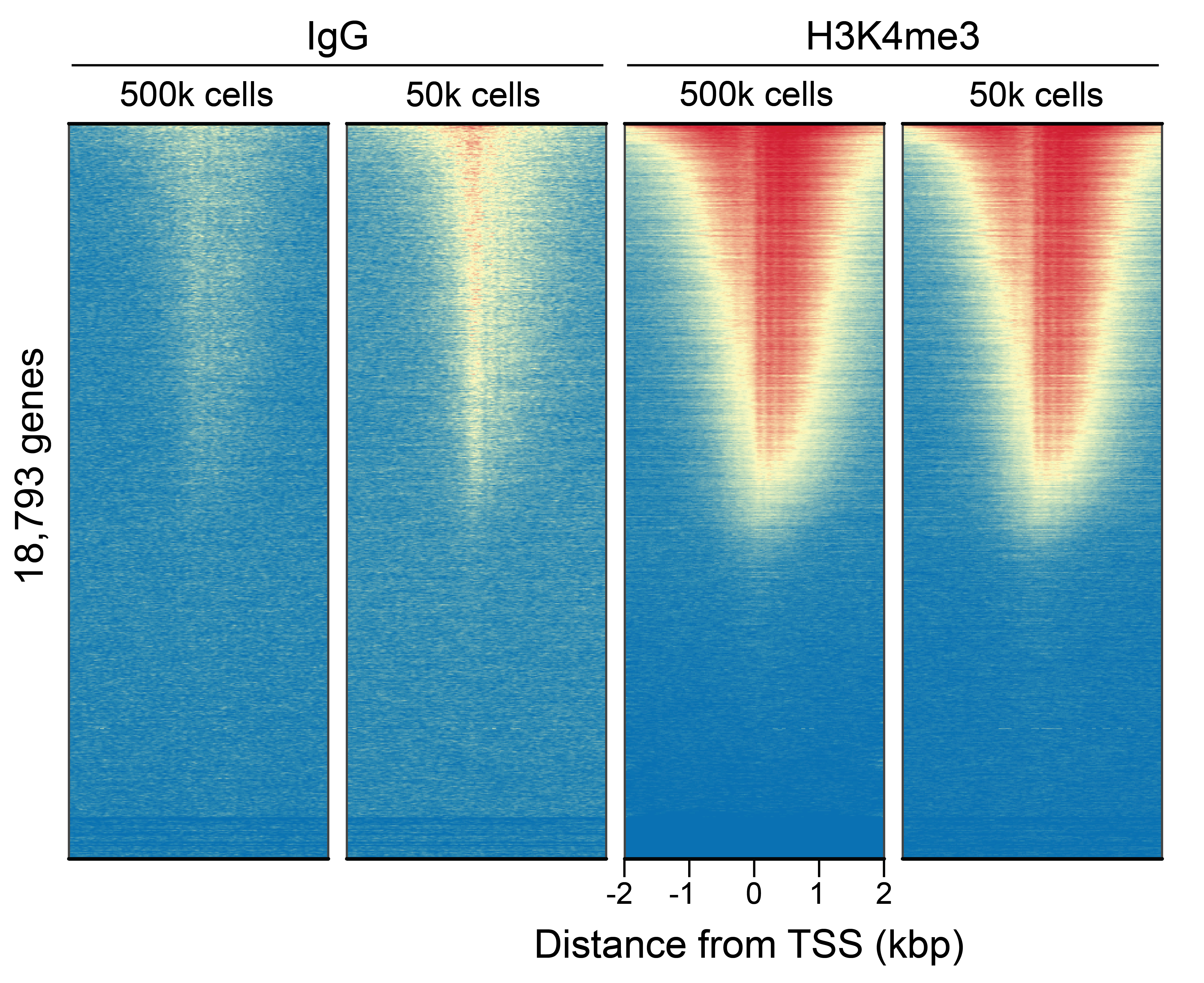
This H3K4me3 (histone H3 lysine 4 trimethyl) antibody meets EpiCypher’s lot-specific SNAP-Certified™ criteria for specificity and efficient target enrichment in CUT&RUN. This requires <20% cross-reactivity to related histone PTMs determined using the SNAP-CUTANA™ K-MetStat Panel of spike-in controls (EpiCypher 19-1002, Figure 1). High target efficiency is confirmed by consistent genomic enrichment at 500k and 50k starting cells (Figures 2-4). This antibody targets histone H3 trimethylated at lysine 4, which is enriched at active promoters near transcription start sites (TSS).
*Mixed Monoclonal: a pool of multiple recombinant monoclonal antibodies.
Validation Data
Figure 1: SNAP specificity analysis in CUT&RUN
CUT&RUN was performed as described in Figure 6. CUT&RUN sequencing reads were aligned to the unique
DNA barcodes corresponding to each nucleosome in the K-MetStat panel (x-axis). Data are
expressed as a percent relative to on-target recovery (H3K4me3 set to 100%).
Figure 2: CUT&RUN genome wide enrichment
CUT&RUN was performed as described in Figure 6. Sequence reads were aligned to 18,793 annotated
transcription start sites (TSSs ± 2 kbp). Signal enrichment was sorted from highest to lowest
(top to bottom) relative to the H3K4me3 - 500k cells sample (all gene rows aligned). High,
medium, and low intensity are shown in red, yellow, and blue, respectively. H3K4me3 antibody
produced the expected TSS enrichment pattern, which was consistent between 500k and 50k cells
and greater than the IgG negative control.
Figure 3: H3K4me3 CUT&RUN representative browser tracks
CUT&RUN was performed as described in Figure 6. Gene browser shots were generated using the
Integrative Genomics Viewer (IGV, Broad Institute). Similar results in peak structure and
location were observed for both cell inputs.
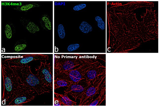
Figure 4: Immunofluorescence
Representative images (60X magnification) of HeLa cells fixed, permeabilized, and immunostained to show endogenous localization of H3K4me3. (A) H3K4me3 antibody (green, 1:100 dilution) detected with an Alexa Fluor® 488 conjugated anti-rabbit secondary. (B) DAPI stained nuclei (blue). (C) Rhodamine stained cytoskeletal F-actin (red). (D) A composite of panels a, b, & c demonstrating nuclear localization of H3K4me3. (E) Negative control lacking H3K4me3 primary antibody to assess background.
Figure 5: Western blot data.
Representative Western data of H3K4me3 in whole cell extracts from HeLa, Hep G2, HCT 116, MCF7, U-2 OS, A549, HEK-293, NIH/3T3, and PC-12 cells. 30 µg of lysate was resolved via SDS-PAGE and detected with H3K4me3 antibody at a 1:250 dilution.
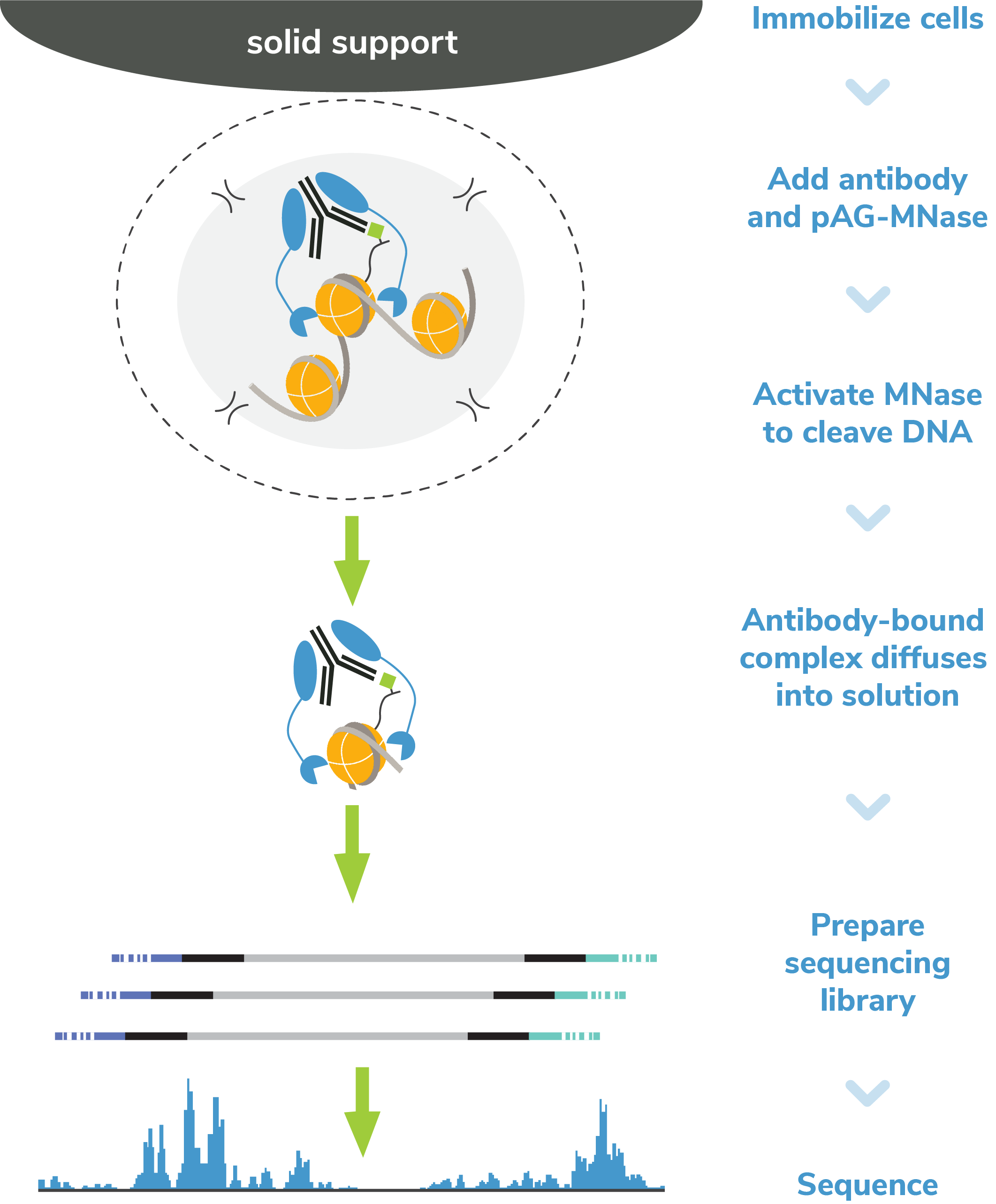
Figure 6: CUT&RUN methods
CUT&RUN was performed on 500k and 50k K562 cells with the SNAP-CUTANA™ K-MetStat Panel (EpiCypher 19-1002) spiked-in prior to the addition of 0.5 μg of either H3K4me3 or IgG negative control (EpiCypher 13-0042) antibodies. The experiment was performed using the CUTANA™ ChIC/CUT&RUN Kit v3 (EpiCypher 14-1048). Library preparation was performed with 5 ng of CUT&RUN enriched DNA (or the total amount recovered if less than 5 ng) using the CUTANA™ CUT&RUN Library Prep Kit (EpiCypher 14-1001/14-1002). Both kit protocols were adapted for high throughput Tecan liquid handling. Libraries were run on an Illumina NextSeq2000 with paired-end sequencing (2x50 bp). Reaction sequencing depth was 8.3 million reads (IgG 500k cell input), 15.5 million reads (IgG 50k cell input), 9.8 million reads (H3K4me3 500k cell input) and 8.6 million reads (H3K4me3 50k cell input). Data were aligned to the hg19 genome using Bowtie2. Data were filtered to remove duplicates, multi-aligned reads, and ENCODE DAC Exclusion List regions.


