CUTANA™ FFPE Transcription Profiling Services
Unlock transcriptional and regulatory insights from archival FFPE tissue
Formalin-fixed, paraffin-embedded (FFPE) tissue archives hold a wealth of clinically relevant biological information, yet conventional methods for investigating transcription and chromatin regulation are often poorly suited to these samples. RNA degradation can compromise RNA-sequencing, while standard ATAC-seq workflows face significant challenges with formalin-fixed tissue.
CUTANA™ FFPE Transcription Profiling Services expand molecular profiling capabilities for archived tissue by mapping RNA Polymerase II occupancy using a DNA-based sequencing readout. Investigate active transcription across protein-coding genes, non-coding RNA genes, and regulatory elements such as enhancers without requiring intact RNA, with EpiCypher managing the workflow from sample processing to sequencing to data delivery.
Ready to Get Started?
Tell us briefly about your project, and a member of our Services team will get back to you shortly!
Unlock More Biology from Archival FFPE Tissue
Access transcriptional insights from aged FFPE
Profile archival FFPE tissue without requiring intact RNA. In our validation across more than 200 samples, blocks up to 30 years old produced high-quality data.
Discover more from less tissue
Access transcriptional and regulatory information using 10-fold less material than FFPE RNA-seq or ATAC-seq. CUTANA™ FFPE only requires a single 5 µm section, conserving valuable specimens for additional analyses. See the data→
Expand your biomarker discovery potential
Explore transcriptional activity across protein-coding genes, non-coding regions, and regulatory elements such as promoters and enhancers to inform biomarker discovery and investigate disease-associated regulatory programs.
Partner with our experts
EpiCypher manages the workflow from tissue processing through sequencing and delivers a tailored data package, allowing your team to stay focused on the biological question.
How Does CUTANA™ FFPE Transcription Profiling Work?
CUTANA™ FFPE Transcription Profiling Services build on FFPE-CUTAC, a published chromatin profiling method developed to map RNA Polymerase II occupancy in formalin-fixed, paraffin-embedded tissue. EpiCypher has further optimized this approach to support an end-to-end transcription profiling service.
Rather than directly measuring mature RNA abundance, the method measures chromatin-associated RNA Polymerase II as a readout of transcriptional activity. Because the assay uses a DNA-based sequencing readout, it does not require intact RNA.
RNA Polymerase II occupancy provides information across actively transcribed genes, including protein-coding and non-coding genes, as well as transcription-associated regulatory regions such as promoters and candidate enhancers. This gives researchers a complementary view of transcriptional biology that is distinct from RNA abundance or chromatin accessibility alone.
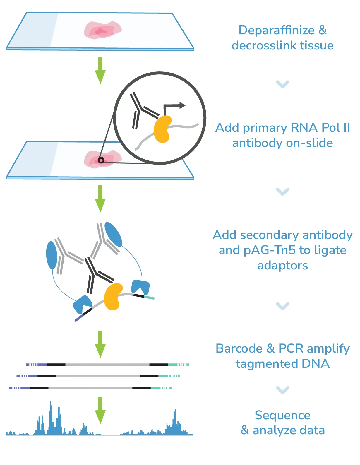
The Science Behind the Service: Building on FFPE-CUTAC
CUTANA™ FFPE Transcription Profiling Services build on published research demonstrating the use of FFPE-CUTAC to recover RNA Polymerase II-associated transcriptional information from fixed tissue. Explore the foundational publications describing FFPE-CUTAC:
Henikoff et al. — Nature Communications, 2023
Foundational description of FFPE-CUTAC for profiling RNA Polymerase II in FFPE samples. View Publication→
Henikoff et al. — Science, 2025
Application of FFPE-CUTAC to understand how hypertranscription might drive cancer. View Publication→
Niu et al. — bioRxiv, 2026 — Preprint
Emerging work benchmarking FFPE-CUTAC against RNA-seq and WGS across archival tumor cohorts. View Preprint→
Explore the Data
CUTANA™ FFPE is designed to turn fixed tissue into biologically interpretable transcriptional information. The following datasets demonstrate performance across transcription-associated genomic features, archival sample ages, and disease-relevant biological programs.
CUTANA™ FFPE Reveals Coding Genes, Non-Coding RNA Genes, and Candidate Enhancers
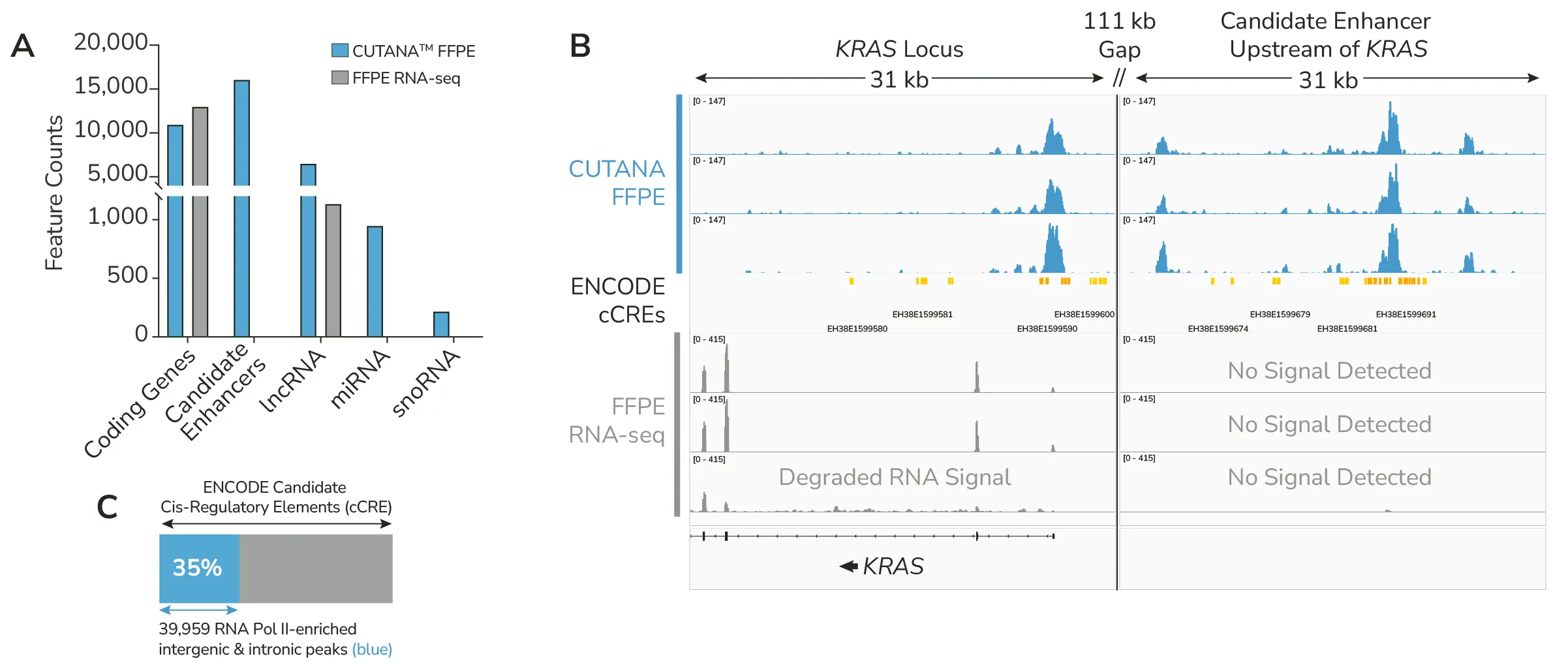
Figure 1. CUTANA™ FFPE detects coding genes, candidate enhancers, and non-coding RNA genes in human lymphoma tissue, while RNA-seq on matched samples detects mainly coding genes. (A) Bar graph detailing the number of genomic features detected by CUTANA™ FFPE and FFPE RNA-seq in matched human lymphoma samples. (B) Representative genome browser tracks for both CUTANA™ FFPE and FFPE RNA-seq at the KRAS locus and an upstream candidate enhancer as cataloged by ENCODE. (C) Proportion bar indicating the overlap between the intergenic and intronic peaks identified by CUTANA™ FFPE in human lymphoma samples and H3K27ac enriched proximal and distal candidate enhancers from the ENCODE database.
FFPE patient lymphoma samples and FFPE RNA-seq data were provided through a collaboration with the laboratory of Dr. Salvia Jain (Massachusetts General Hospital Cancer Center), with Drs. Makoto Iwasaki and Eric Minwei Liu contributing to study design, experimentation, analysis and biological interpretation. Genome browser tracks were compared to Version 2 of the ENCODE registry of cCREs: ENCODE Project Consortium, Jill E. Moore, Michael J. Purcaro, Henry E. Pratt, Charles B. Epstein, Noam Shoresh, Jessika Adrian, et al. 2020. “Expanded Encyclopaedias of DNA Elements in the Human and Mouse Genomes.” Nature 583 (7818): 699–710.
Profile Years-Old Archival FFPE With Strong Transcription-Associated Signal
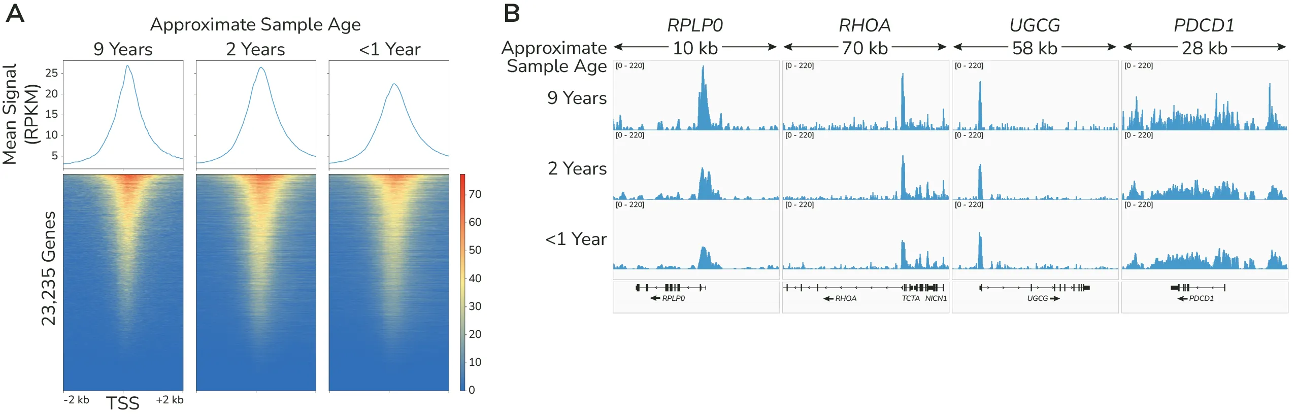
Figure 2. CUTANA™ FFPE shows strong transcription-associated signal across samples of various ages. (A) Transcription start site (TSS) enrichment plots for 9-, 2-, and <1-year-old human FFPE samples from lymphoma patients. Heatmap rows are sorted by signal intensity in the 9-year-old sample and held constant across panels for direct comparison. (B) Representative genome browser tracks at the same 9-, 2-, and <1-year-old human lymphoma samples at the housekeeping gene RPLP0 and the lymphoma-associated loci, RHOA, UGCG, and PDCD1.
FFPE patient lymphoma samples and FFPE RNA-seq data were provided through a collaboration with the laboratory of Dr. Salvia Jain (Massachusetts General Hospital Cancer Center), with Drs. Makoto Iwasaki and Eric Minwei Liu contributing to study design, experimentation, analysis and biological interpretation.
Achieve Strong TSS Enrichment in FFPE Where Standard ATAC-seq Falls Short
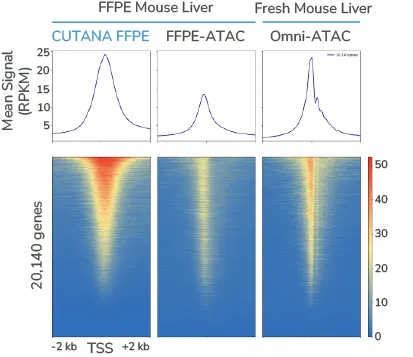
Figure 3. CUTANA™ FFPE delivers higher signal intensity than ATAC-based methods in mouse liver tissue. Transcription start site (TSS)-centered heatmaps comparing signal in mouse liver across 20,140 annotated genes. CUTANA™ FFPE showed the highest overall signal, followed by Omni-ATAC, and substantially lower signal in FFPE-ATAC.
Comparison ATAC-seq data sourced from: FFPE-ATAC (FFPE liver): SRRJ 5328707 [Zhang et al., Genome Res 2022. PMID: 34261731]. Omni-ATAC (normal liver): SRR8119838 [Liu et al., Sci Data 2019. PMID: 31110271]. Heatmaps are row linked and show signal sorted relative to the CUTANA FFPE sample.
Recover Biologically Relevant Transcriptional Programs from Fixed Tissue
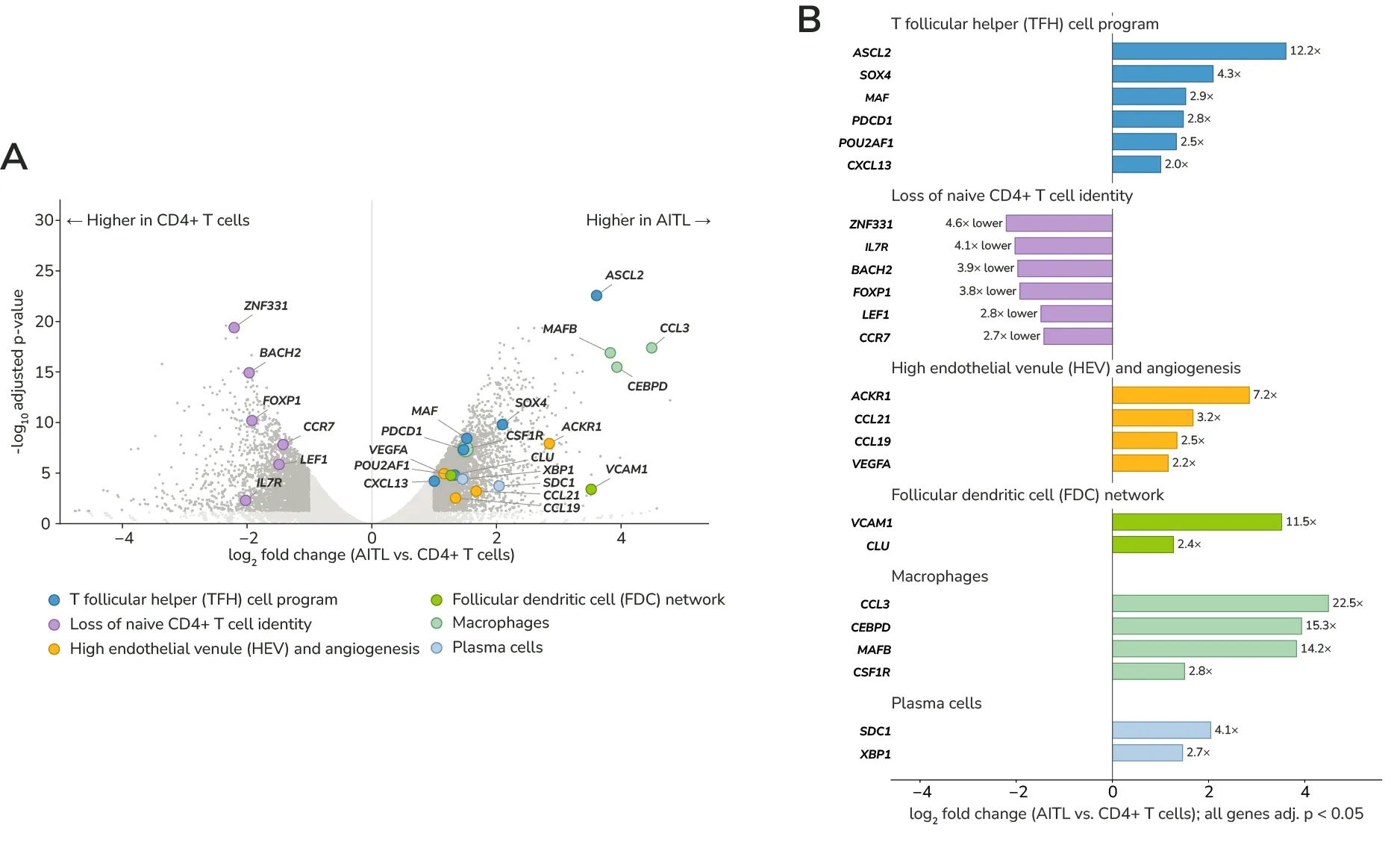
Figure 4. CUTANA™ FFPE identifies disease-associated transcriptional programs in archived human lymphoma tissue. CUTANA™ FFPE signal was analyzed across gene bodies (from transcription start site to transcription termination site) for three angioimmunoblastic T-cell lymphoma (AITL) biological replicates and three normal CD4+ T cell controls. (A) Volcano plot highlighting the significant changes in RNA Pol II engagement (adjusted p-value < 0.05, ≥ 2-fold change) based on differential analysis. (B) Bar graph representing the fold change for selected significant genes related to the pathogenesis of AITL.
FFPE patient lymphoma samples and FFPE RNA-seq data were provided through a collaboration with the laboratory of Dr. Salvia Jain (Massachusetts General Hospital Cancer Center), with Drs. Makoto Iwasaki and Eric Minwei Liu contributing to study design, experimentation, analysis and biological interpretation.
How Does Your Fully Managed Project Work?
Discuss your samples and research question
Partner with EpiCypher scientists to evaluate your project goals, available tissue, and experimental strategy.
Send your FFPE tissue
Ship your FFPE tissue sections on positively charged slides for processing by our Genomic Services team.
We perform the CUTANA™ FFPE assay and sequencing
EpiCypher manages tissue processing, CUTANA™ FFPE profiling, and sequencing.
Receive your tailored data package
Every project concludes with a one-on-one review of your results, delivered as a conversation with our experts.
FAQs About Our CUTANA™ FFPE Transcription Profiling Services
What is FFPE-CUTAC?
FFPE-CUTAC is the published chromatin profiling method underlying CUTANA™ FFPE Transcription Profiling Services. The method was developed to map RNA Polymerase II occupancy in formalin-fixed, paraffin-embedded material. EpiCypher has further optimized this approach to support an end-to-end transcription profiling service.
Is CUTANA™ FFPE the same as RNA-seq?
No. RNA-seq measures RNA abundance, while CUTANA™ FFPE profiles chromatin-associated RNA Polymerase II occupancy. These readouts provide related but distinct information about transcription.
How is CUTANA™ FFPE different from ATAC-seq?
ATAC-seq profiles chromatin accessibility genome-wide, while CUTANA™ FFPE maps chromatin-associated RNA Polymerase II to provide insights into transcriptional activity and chromatin accessibility at transcription-coupled regions. CUTANA™ FFPE delivers strong transcription start site (TSS) enrichment from a single 5 µm FFPE tissue section, using approximately 10-fold less tissue than reported FFPE-adapted ATAC-seq methods.
How much material do I need for a CUTANA™ FFPE service project?
For a CUTANA™ FFPE Service project you should submit one anonymized 5 µm section per sample. The tissue area of a section should be approximately 1 cm2. If you have any questions on whether your amount of material is sufficient for CUTANA™ FFPE Services, contact us to discuss your project with our experts.
What if the area of my tissue section is less than 1 cm²?
The success of your sample depends on both the area of the material provided and the cellularity of the tissue. We have observed the most consistent success with tissues that reach the minimum 1 cm2 threshold but we have observed success with smaller tissue sections (down to 2 mm2) that maintain high levels of cellularity. Submitting a smaller tissue amount will result in a higher risk of failure for your FFPE sample. If you have any questions about the suitability of your sample for CUTANA™ FFPE, contact us to discuss your project with our experts.
Can I submit serial tissue sections?
If your serial sections both fit within the assay area (28 mm x 17 mm) in the center of a charged microscopy slide then both sections can contribute to the 1 cm2 tissue area recommendation for this assay.
What fixation conditions are compatible with CUTANA™ FFPE?
Many archival samples were prepared before any downstream molecular assay was planned, so variation in fixation is common and does not automatically rule a sample out. CUTANA™ FFPE was optimized for human and mouse FFPE material fixed in either 10% neutral buffered formalin or 4% paraformaldehyde at a 1:20 tissue-to-fixative ratio, for 12–24 hours at room temperature. Additionally, we recommend ischemia time to be kept to under 3 hours. Deviations from these conditions can affect results to different degrees:
- Extended fixation: DNA quality declines progressively with time in fixative, most noticeably beyond 2–3 days. Samples fixed for up to 72 hours are generally acceptable; longer fixation increases the risk of lower signal.
- Unbuffered or acidic formalin: this causes substantially more DNA damage than neutral buffered formalin and is the variable most likely to compromise results.
- Prolonged delay to fixation: delays of 3 hours or more introduce more artefacts into genome-wide analyses.
If you have any questions about the fixation conditions of your samples, contact us to discuss your project.
Can I submit stained FFPE sections?
We ask that you submit unstained, paraffin embedded sections for profiling. Staining procedures, cover-slipping, and mounting media can interfere with the CUTANA™ FFPE workflow and prevent us from processing your sample. We do encourage you to perform H&E staining of an adjacent section to confirm tissue quality and cellularity, but that section cannot be used for CUTANA™ FFPE profiling.
How can I assess the cellularity of my samples?
The quickest way to assess cellularity is to review an H&E-stained section cut from the same block. Hematoxylin binds nucleic acids and stains nuclei dark blue-purple, while eosin binds cytoplasmic and extracellular proteins and stains cytoplasm and stroma in shades of pink. This contrast makes cellularity easy to estimate by eye:
- High cellularity: densely packed, dark purple nuclei with little intervening pink space.
- Low cellularity: sparse, widely spaced nuclei against a predominantly pink background.
Your pathology core or a pathologist can also provide an estimate of percentage cellularity or tumour content if needed. If your H&E shows low cellularity it may impact the data quality of your sample.
How can I tell whether my FFPE samples are suitable for CUTANA™ FFPE profiling??
FFPE material is highly variable, and no single measurement guarantees success, but several pre-analytical factors are strong predictors. Samples are most likely to succeed when they:
- Have been fixed in neutral buffered formalin for 12–24 hours;
- Had a short delay to fixation (under 3 hours);
- Show adequate cellularity on H&E with intact tissue morphology;
- Have been successfully profiled using molecular assays such as RNA-seq or DNA-seq.
Block age alone is not a reliable predictor: in our validation across more than 200 samples, blocks up to 30 years old produced good quality data, with an overall success rate above 75%. Tissue type and pre-analytical handling generally matter more.
For important or limited cohorts, we recommend a pilot with a small number of representative samples before committing the full set.
Ask our team if you have any questions!
What data do I receive?
CUTANA™ FFPE is a modified CUT&Tag workflow that maps RNA Polymerase II enrichment across the genome directly in FFPE tissue sections. You receive genome-wide profiles showing where RNA pol II is transcriptionally active at promoters and across gene bodies, as well as, RNA pol II binding to regulatory regions including candidate enhancers, lnRNAs, miRNAs, snoRNAs, and small non-polyadenylated genes such as histones.
Standard deliverables for this service include:
- Raw sequencing reads (FASTQ files).
- Aligned reads (BAM files) mapped to the appropriate reference genome.
- Normalized signal tracks (bigWig files) for viewing in genome browsers such as IGV or UCSC.
- Peak calls (BED files) identifying enriched regions.
- A QC report summarizing sequencing depth, alignment rate, duplication and enrichment metrics for each sample.
How do I analyze my CUTANA™ FFPE data?
- Epigenomic and regulatory analysis: Similar to ATAC-seq, ChIP-seq, CUT&RUN, and CUT&Tag, this approach uses peak calling to identify regions of enrichment to investigate gene regulation, including promoter and enhancer activity.
- Transcriptional profiling: Using an analysis approach similar to RNA-seq, we quantify RNA Polymerase II enrichment across annotated genes to investigate transcriptional regulation, identify differences between conditions, and characterize affected biological pathways.