Single-Cell CUT&Tag: pushing the boundaries of epigenomic profiling
- Nathaniel Wesley
CUT&Tag is a novel epigenomic mapping technology that builds on the related sister chromatin-immunotethering technology CUT&RUN. CUT&Tag generates high-quality data with ultra-low cell inputs using a fraction of the sequencing depth compared to traditional ChIP-seq. In this blog post, we outline the development of CUT&Tag and describe its adoption for single-cell experiments. Single-cell CUT&Tag (and CUT&RUN) is poised to transform our understanding of epigenetic regulation and disease, particularly through analysis of complex inputs such as heterogeneous tissues and patient samples.
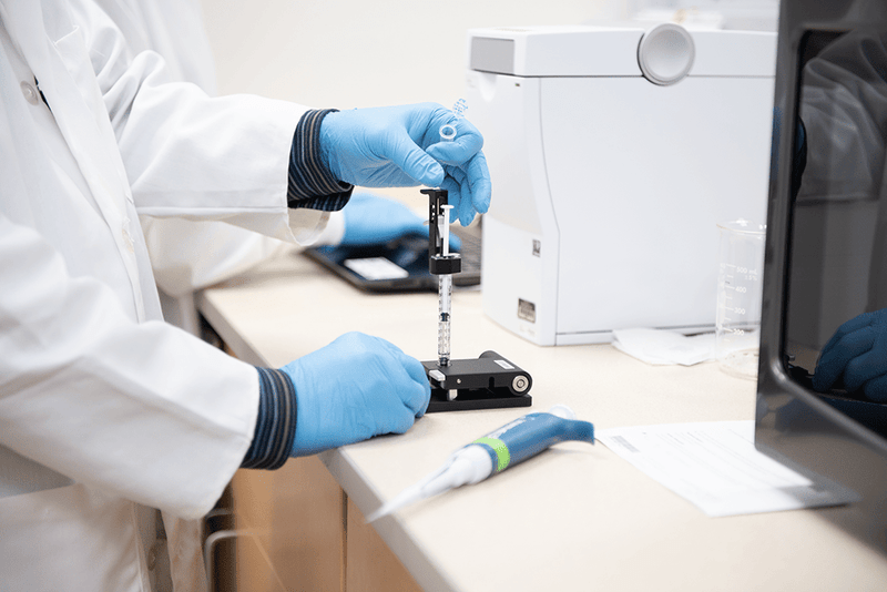
If you are interested in designing your own CUT&Tag experiment, view the EpiCypher CUTANA™ CUT&Tag product overview where you can find reagents as well as our optimized CUT&Tag protocol. Also check out our introduction to CUT&Tag, which covers the basics and links to additional helpful information.
Evolution of Chromatin Immunotethering Methods
Genome templated processes occur in a milieu of transcription factors, chromatin-associated proteins and histone post-translational modifications (PTMs). Characterizing this complex regulatory network is essential for understanding fundamental biological processes and their misregulation in disease. Chromatin immunoprecipitation sequencing (ChIP-seq) was a landmark development in the field and yielded critical insights about the distribution and abundance of epigenetic targets1. However, the basic protocol remains unchanged over 35 years later, and suffers from low signal-to-noise, large cell input requirements, and high costs, all of which make this technique impractical for clinical projects.
Dr. Steven Henikoff’s laboratory has leveraged recent innovations in immunotethering technology2 to develop streamlined, “ChIP-less” chromatin profiling approaches. In each assay, cells (or nuclei) are immobilized to a solid support, permeabilized, and treated with a target-specific antibody. An enzyme is then tethered to antibody-labelled chromatin via a protein A/G (pAG) fusion to direct enzymatic activity towards these regions of interest.
- CUT&RUN (Cleavage Under Targets and Release Using Nuclease) uses a pAG-micrococcal nuclease (MNase) fusion to selectively cleave antibody-bound chromatin. Fragmented DNA is released into solution, purified, and prepared for next-generation sequencing (NGS)3. You can read about the development of CUT&RUN and several recent innovative applications of this technology in other EpiCypher blog posts.
- CUT&Tag (Cleavage Under Targets and Tagmentation) applies a pAG-Tn5 transposase to simultaneously cleave and “tag” antibody-bound chromatin with sequencing adaptors. The tagmented DNA is selectively amplified via PCR and used for NGS4,5.
By selectively targeting chromatin regions of interest, both CUT&RUN and CUT&Tag generate data with exquisite signal-to-noise using ~10-fold fewer sequencing reads vs. ChIP-seq. This enables epigenomic profiling using significantly less input material, with both CUT&RUN6,7 and CUT&Tag4 pushing the boundaries to single cell profiling.
So which assay is better – CUT&Tag or CUT&RUN? It depends!
CUT&RUN and CUT&Tag are complementary assays, and each brings distinct advantages in certain contexts. The use of Tn5 in CUT&Tag has drawn comparisons to ATAC-seq (Assay for Transposase-Accessible Chromatin), which relies on the non-specific preference of Tn5 activity towards open chromatin to map accessibility from extremely low cell inputs8. Direct integration of sequencing adapters by Tn5 eliminates time-intensive and costly library prep steps required for traditional ChIP-seq and CUT&RUN. Fewer steps also reduces risk of sample loss – key for single cell sequencing applications.
Since its introduction in 2013, ATAC-seq has been optimized for both single-cell mapping and automated workflows8,9, demonstrating the compatibility of tagmentation techniques with advanced epigenomic analyses. Although “ATAC-like” signal is a concern in CUT&Tag experiments, the high salt wash steps help reduce nonspecific pAG-Tn5 binding. However, this assay modification comes at a cost – low abundant or weakly associated chromatin proteins may be displaced by high salt washes. Furthermore, because Tn5 activity is intrinsically biased towards regions of open chromatin, CUT&Tag may not be optimal for studying repressive marks.
Thus, CUT&Tag is best suited for mapping histone PTMs and some abundant transcription factors associated with accessible chromatin, especially when working with reduced inputs. CUT&RUN, in contrast, is robust for diverse targets, including transcription factors, chromatin remodeling enzymes and histone PTMs, as well as various cell types and input amounts. Of note, the CUT&RUN assay is not readily compatible with scalable single cell analysis workflows, such as droplets or combinatorial indexing. Knowing these factors, EpiCypher recommends CUT&RUN for most bulk analyses and CUT&Tag for low input studies on histone PTMs.
Single-Cell CUT&Tag
Due to their high sensitivity, it is no surprise that CUT&Tag4, CUT&RUN6,7,10, and ATAC-seq9,11–13 have been adapted for single-cell analyses. Here we will focus on the single-cell applications of CUT&Tag, which have attracted many researchers due to the use of Tn5 tagmentation technology.
Perhaps the most powerful feature of CUT&Tag is that the entire reaction – from antibody binding to tagmentation of sequencing adapters – occurs within intact nuclei (or cells). Thus, creation of a single cell suspension following a bulk CUT&Tag experiment facilitates barcoding of individual cells via indexing PCR. The resulting libraries can then be pooled for multiplex sequencing, making CUT&Tag a highly attractive strategy for single-cell chromatin profiling.
In the original CUT&Tag paper, Kaya-Okur et al. used a Takara iCell8 nano-dispensing system to isolate over 1,000 individual cells for high-throughput single-cell CUT&Tag (scCUT&Tag) experiments4. Importantly, they demonstrated that scCUT&Tag data for H3K4me2 and H3K27me3 correlate with profiles from corresponding bulk cell populations. The resulting single cell histone PTM maps could also be used to distinguish between H1 and K652 cell types in silico with high accuracy.
Bartosovic et al. adapted the scCUT&Tag protocol to investigate histone PTMs and transcription factor occupancy in single cells isolated from mouse brain tissue14. Using scCUT&Tag they were able to distinguish unique cell types and characterize changes in histone PTMs (H3K4me3, H3K27ac, H3K36me3, and H3K27me3) throughout cell differentiation. They also applied scCUT&Tag to map the transcription factor OLIG2 and the cohesin complex component RAD51, low-abundant targets that have been challenging to profile at single-cell resolution. These results reveal the epigenetic heterogeneity within tissue samples and establish the benefit of single-cell epigenomics, despite technical challenges and sparse data compared to bulk methods.
Zhu et al. combine scCUT&Tag with RNA-seq to simultaneously profile five histone PTMs (H3K4me1/3, H3K27ac, H3K27me3, and H3K9me3) and gene expression in single cells isolated from mouse brain tissue15. The authors demonstrate the power of “multiomics” approaches where the integrated analysis of transcriptional activity, histone modifications, and chromatin accessibility (via Paired-seq16) can uncover the intricate epigenetic regulatory mechanisms governing different cell types.
Clinical Applications of CUT&Tag
Single-cell analysis has the potential to uncover epigenetic programs driving development and human disease at unprecedented resolution. Janssens et al. implemented a CUT&Tag strategy to profile heterogeneity in mixed-lineage leukemia (MLL) patient samples, healthy patient samples, and cell lines 17. Specifically, the authors investigated the H3K4 methyltransferase KMT2A, which is translocated in approximately 10% of acute leukemias and has been shown to fuse with transcriptional elongation factors (e.g. AF9, AF4). Although it is known that different KMT2A rearranged oncofusion proteins (referred to as KMT2Ar for brevity) have distinct genomic localization, it has been difficult to accurately map and characterize their function due to limited patient samples and the poor resolution of existing approaches.
To enable efficient mapping from small patient samples, Janssens et al. developed automated CUT&Tag assays, and used them to profile various KMT2Ar fusion proteins and a collection of histone PTMs denoting active and silenced chromatin. They identified a subset of KMT2Ar binding sites with bivalent (H3K4me3 + H3K27me3) chromatin signatures. They then used scCUT&Tag to determine that this bivalent signature in the bulk population was caused by heterogeneity among individual cells within the tumor.
Finally, the authors used automated CUT&Tag to characterize cellular responses and predict tumor sensitivity to epigenetic inhibitors. DOT1L is highly indicated in cancer development and has been shown to interact with KMT2Ar fusion proteins in some leukemias. CUT&Tag provides the sensitivity needed to detect differential recruitment of DOT1L by KMT2Ar and monitor the impact of DOT1L inhibitors in various leukemia cell lines. Although not yet at single-cell resolution, this approach demonstrates CUT&Tag as a robust platform to map patient-specific fusion proteins, compare with profiles of other important factors or histone PTMs, and subsequently use this information to rationally determine best courses of treatment.
Conclusion
CUT&RUN and CUT&Tag are revolutionizing epigenomics research through economical high-throughput profiling of histone PTMs, transcription factors, and other chromatin-associated proteins. The development of automated CUT&RUN and CUT&Tag assays will help standardize both single cell and bulk population profiling, while also providing the sensitivity required for clinical research projects. scCUT&Tag in particular has the potential to support clinical epigenetics through evaluation of rare or limited patient samples to guide personalized treatment courses.
EpiCypher is at the forefront of novel epigenomics technologies. We have rigorously validated antibodies and reagents to support your CUT&Tag workflow, including the critical pAG-Tn5 enzyme. We also recommend incorporating our newly-released SNAP-CUTANA™ Spike-in Controls, panels of DNA-barcoded modified nucleosomes which enable rigorous assay development, antibody validation, sample normalization, and quantitative cross-sample comparisons. Contact us at [email protected] for more information!
Follow us on Twitter and LinkedIn for the latest news in chromatin biology!
References
- Solomon, M. J., and Varshavsky, A. Formaldehyde-mediated DNA-protein crosslinking: A probe for in vivo chromatin structures. PNAS 82, 6470–6474 (1985). PubMed PMID: 2995966.
- Schmid, M. et al. ChIC and ChEC: Genomic Mapping of Chromatin Proteins. Mol. Cell 16, 147–157 (2004). PubMed PMID: 15469830.
- Skene, P. J., and Henikoff, S. An efficient targeted nuclease strategy for high-resolution mapping of DNA binding sites. ELife 6, 1–35 (2017). PubMed PMID: 28079019.
- Kaya-Okur, H. S., et al. CUT&Tag for efficient epigenomic profiling of small samples and single cells. Nat. Commun. 10, 1–10 (2019). PubMed PMID: 31036827.
- Kaya-Okur, H. S., et al. Efficient low-cost chromatin profiling with CUT&Tag. Nat. Protoc. 15, 3264–3283 (2020). PubMed PMID: 32913232.
- Hainer, S. J., et al. Profiling of Pluripotency Factors in Single Cells and Early Embryos. Cell 177, 1319-1329.e11 (2019). PubMed PMID: 30955888.
- 7. Patty, B. J., and Hainer, S. J. Transcription factor chromatin profiling genome-wide using uliCUT&RUN in single cells and individual blastocysts. Nat. Protoc. 16, 2633–2666 (2021). PubMed PMID: 33911257.
- Buenrostro, J. D., et al. Transposition of native chromatin for fast and sensitive epigenomic profiling of open chromatin, DNA-binding proteins and nucleosome position. Nat. Methods 10, 1213–1218 (2013). PubMed PMID: 24097267.
- Cusanovich, D. A., et al. The cis-regulatory dynamics of embryonic development at single-cell resolution. Nature 555, 538–542 (2018). PubMed PMID: 29539636.
- Ku, W. L., et al. Single-cell chromatin immunocleavage sequencing (scChIC-seq) to profile histone modification. Nat. Methods 16, 323–325 (2019). PubMed PMID: 30923384.
- Cusanovich, D. A., et al. Multiplex single-cell profiling of chromatin accessibility by combinatorial cellular indexing. Science 348, 910–914 (2015). PubMed PMID: 25953818.
- Cao, J., et al. Joint profiling of chromatin accessibility and gene expression in thousands of single cells. Science 361, 1380–1385 (2018). PubMed PMID: 30166440.
- Vitak, S. A., et al. Sequencing thousands of single-cell genomes with combinatorial indexing. Nat. Methods 14, 302–308 (2017). PubMed PMID: 28135258.
- Bartosovic, M., et al. Single-cell CUT&Tag profiles histone modifications and transcription factors in complex tissues. Nat. Biotechnol. 39, 825-835 (2021). PubMed PMID: 33846645.
- Zhu, C., et al. Joint profiling of histone modifications and transcriptome in single cells from mouse brain. Nat. Methods 18, 283–292 (2021). PubMed PMID: 33589836.
- Zhu, C., et al. An ultra high-throughput method for single-cell joint analysis of open chromatin and transcriptome. Nat. Struct. Mol. Biol. 26, 1063–1070 (2019). PubMed PMID: 31695190.
- Janssens, D. H., et al. Automated CUT&Tag profiling of chromatin heterogeneity in mixed-lineage leukemia. Nat. Genet. 53, 1586-1596 (2021). PubMed PMID: 34663924.